0335
Accelerating perfusion quantification using ASL-MRI with a neural network based forward model1Institute of Biomedical Engineering, Department of Engineering Science, University of Oxford, Oxford, United Kingdom, 2Sir Peter Mansfield Imaging Centre, School of Medicine, University of Nottingham, Nottingham, United Kingdom, 3Wellcome Centre for Integrative Neuroimaging, FMRIB, Nuffield Department of Clinical Neurosciences, University of Oxford, Oxford, United Kingdom
Synopsis
Arterial Spin Labelling is established as a quantitative technique to measure perfusion and other hemodynamic properties of the cerebral vasculature. This application of ASL requires multiple post label delays and parameter estimation via a kinetic model. However, the computational cost of the post-processing can be an issue, especially with sophisticated kinetic models. In this work, we propose a rapid method to perform perfusion estimation by replacing kinetic models with pre-trained neural networks. Two neural networks were trained to replace the kinetic model with or without gamma dispersion effects. The dispersion neural network is shown to achieve a lower computational cost.
Introduction
Arterial spin labeling (ASL) is a magnetic resonance imaging (MRI) technique designed for quantifying absolute perfusion values1. Perfusion quantification relies on the theory of tracer kinetics, the relationship between signal and perfusion is modeled as a mathematical formula, which is then used to convert from signal intensity to measured perfusion. Currently, the standard approach is to use the Buxton kinetic model (KM) to describe this relationship2. To increase the accuracy of ASL perfusion measurements, adjustments may be applied to the model to account for factors such as dispersion effects3 or macrovascular contamination4, but this comes at a cost of model complexity. Application of the model to measured data requires an ‘inversion’ of the model, for multi-time point (e.g., multi post label delay) data some form of model fitting is required and Bayesian inference strategies have been employed that offer both parameter estimates and their associated uncertainty5. Existing model-fitting and Bayesian inference algorithms reply on iterative algorithms that require multiple evaluations of the kinetic model. The computational cost in estimating perfusion consequently grows as the complexity of forward model increases. To improve the computational efficiency, in this work we propose to replace direct evaluation of the KM by pre-trained neural networks (NN) within a Bayesian inference algorithm. In the previous literature, the use of NN to directly estimate perfusion from ASL does not provide uncertainty information and does not generalize. Our work achieves the uncertainty estimation and generalizability by Bayesian inference and the careful design of NN.Methods
In this work, we trained two fully connected multilayer perceptrons (MLP), NNgeneral and NNdispersion, to replace the KM with or without gamma dispersion effects3. Each neural network contained three fully connected layers with 10 neurons in each layer. In each case we sampled 2,000,000 synthetic data points from the corresponding kinetic model as the training data. ReLu function, 5-fold cross-validation technique and Adam optimizer with initial learning rate = 0.001, β1 = 0.9, β2 = 0.9.In order to retain the generalizability of having a full KM, in training the first MLP to replace the standard KM, arterial transit time (ATT), relaxation constant of tissue (T1), relaxation constant of blood (T1b), label duration (τ) and inflow time (t) were taken as the inputs with corresponding training values ranging from 0 to 5 covering the recommended labeling values and physiological plausible ranges according to ASL consensus paper1.The second MLP contained two more inputs, the log scales of sharpness (s) and time-to-peak (p) with training ranges between (1,3) and (-3,-1). Since the KM is approximately linear with perfusion this was not included in the NN and a unity scaled KM was learned with the output being scaled by the perfusion in the model inference algorithm. The training process was performed in python using the scikit-learn library.
We conducted experiments estimating perfusion and ATT together on both simulated data and in-vivo data with KMs or NNs as the model in a stochastic variational Bayesian inference algorithm6. Data were simulated with perfusion of 60 ml/100g/min and ATT varying between 0 and 2 seconds, every 0.05 seconds. The acquisition and physiological parameters were specified to the recommended values from consensus paper1 with τ = 1.8, T1 = 1.3 and T1b = 1.65. The PLDs were chosen as CBF-ATTopt proposed by Woods7. Gaussian noise was added to generate data with SNR of infinity, 10, 5 and 2.5. In each case, 100 repetitions were generated. We compared the average computational time, the estimated perfusion values and estimated ATT between the cases with KM or NN as the forward model. The in-vivo data was acquired using PCASL from a single individual. The dataset contained a total of 96 measurements which included 6 PLDS (0.25, 0.5, 0.75, 1.0, 1.25, 1.5) with each repeated eight times.
Results
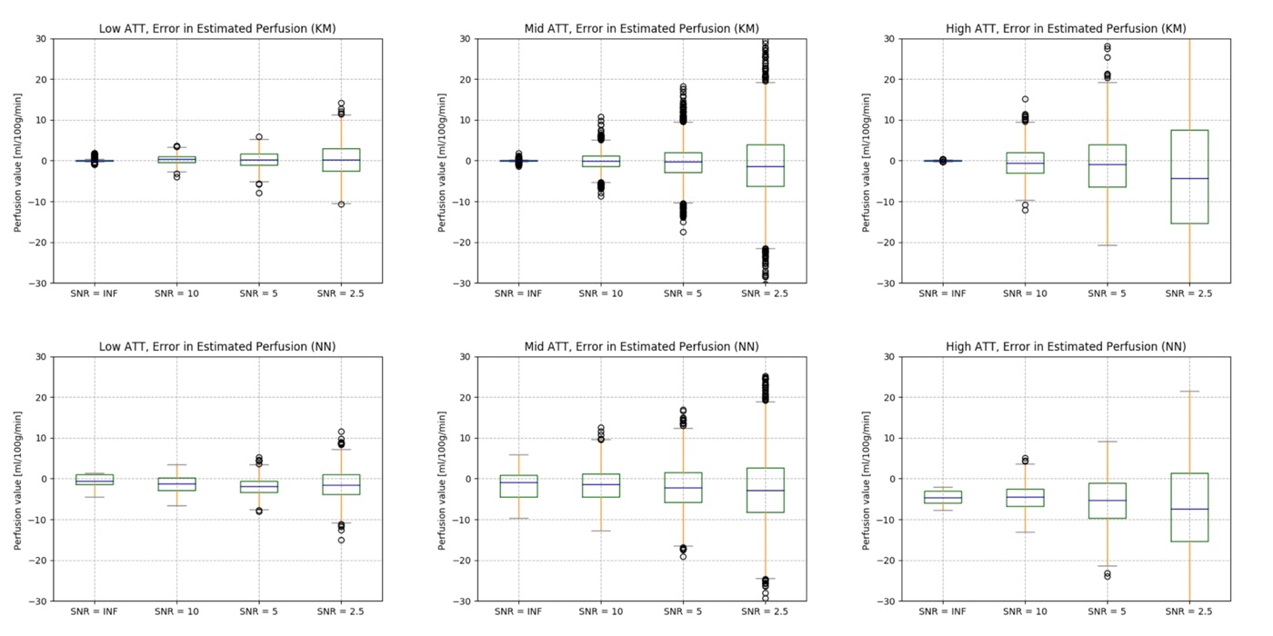
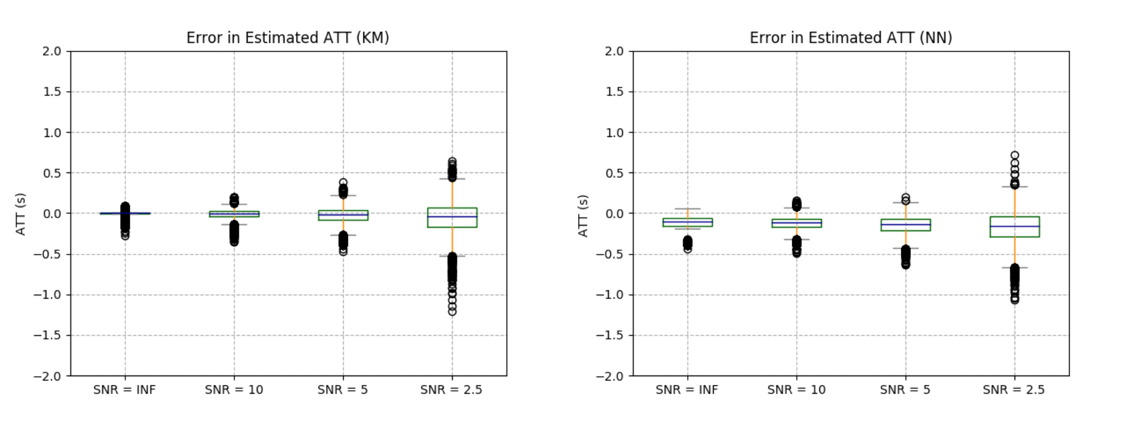
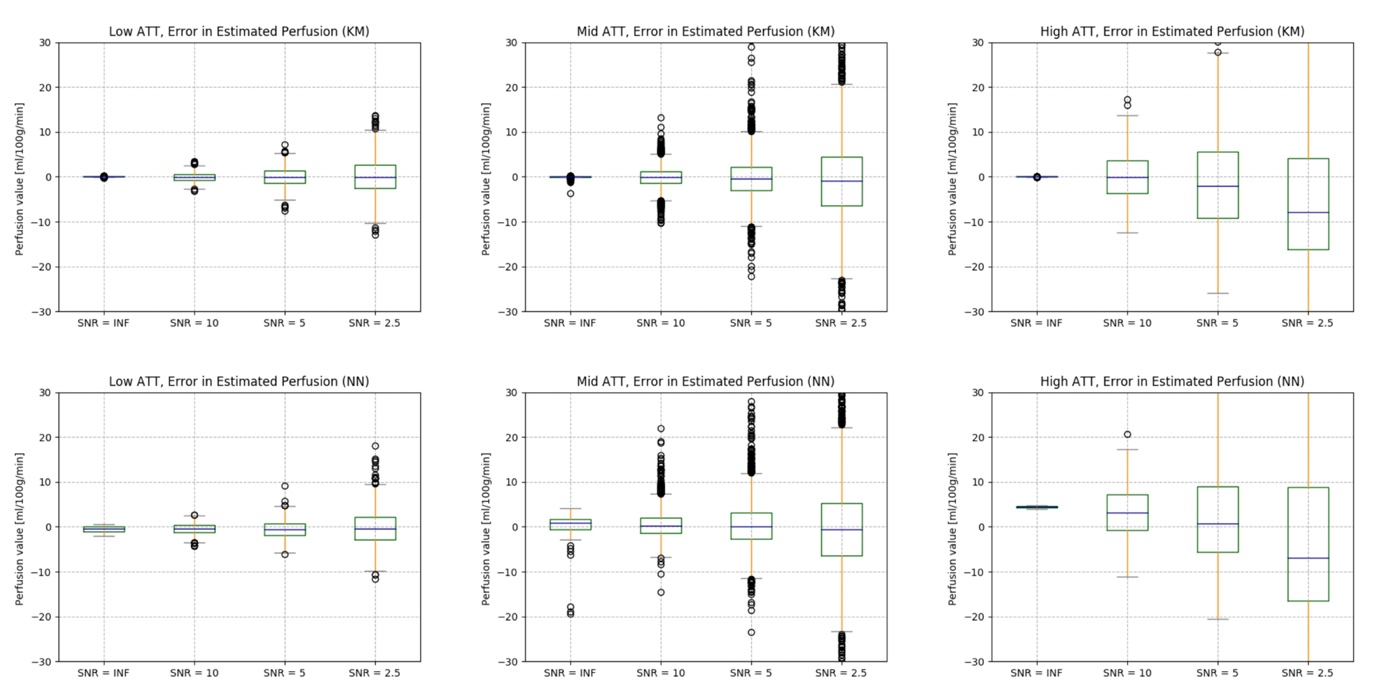
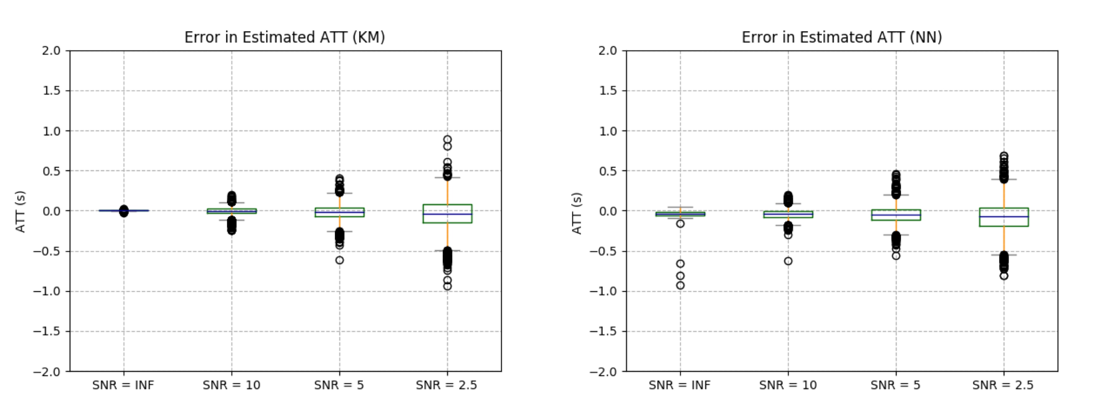
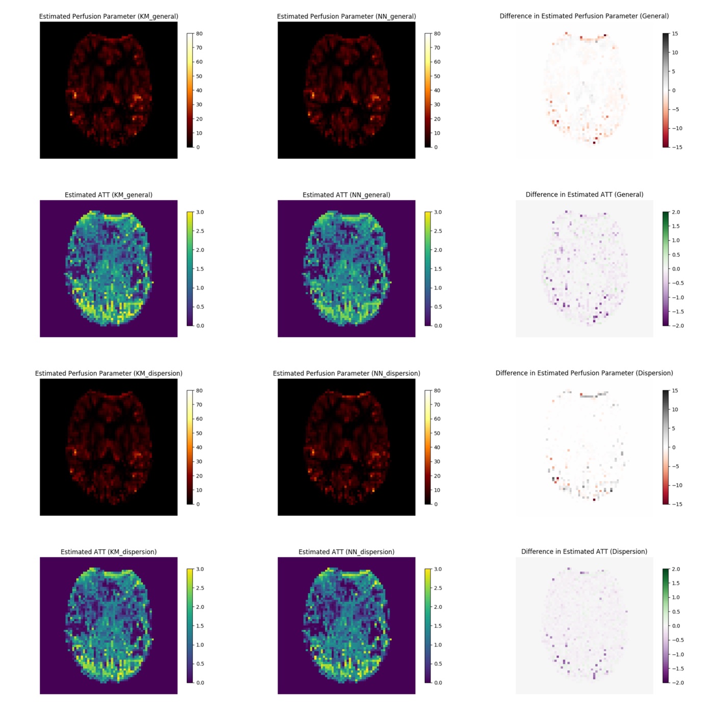
Figure 1 and 2 provide the comparisons between the general kinetic model and NNgeneral in simulation experiments. Both models performed well but NNgeneral exhibited slightly more bias in perfusion and ATT errors while the variance was consistent. The average computational time for each simulation experiment was found to be 31.15s (NNgeneral) versus 29.45s (general KM). Figure 3 and 4 show the comparisons between the dispersion KM and NNdispersion in simulation experiments. Both models performed well with little overall bias for all cases and the variance was consistent. The computational time for simulation experiment was 26.64s (NNdispersion) versus 147.10s (gamma dispersion KM), indicating a 5-fold improvement. Figure 5 shows the results from in-vivo experiments, suggesting a similarity between NN-based and KM-based approaches.Conclusion and Discussion
The ultimate goal of our project was to build up an efficient and uncertainty-aware parameter estimation framework. As the first attempt, we replaced ASL KMs with NNs within the inference algorithm. Our simulation results indicated a 5-fold improvement in computational cost by replacing gamma dispersion KM by a NN without substantial loss in accuracy. The benefits of this approach are likely to be even greater when used with more complex non-linear models.Acknowledgements
This research was supported by Institute of Biomedical Engineering, Department of Engineering Science, University of Oxford. I am grateful to my supervisor Prof Michael Chappell who provided guidance and feedback throughout this project. I am also thankful to my colleague Martin Craig for the assistance with coding and the other colleagues at my group for their comments on earlier versions of manuscript.References
1. David C Alsop, John A Detre, Xavier Golay, G Matthias, Jeroen Hendrikse,Luis Hernandez-garcia, Hanzhang Lu, Bradley J Macintosh, Laura M Parkes,Marion Smits, Matthias J P Van Osch, Danny J J Wang, Eric C Wong, andGreg Zaharchuk. Recommended Implementation of Arterial Spin-LabeledPerfusion MRI for Clinical Applications : A Consensus of the ISMRM PerfusionStudy Group and the European Consortium for ASL in Dementia.116(October 2013):102-116, 2015
2. Buxton, R. B., Frank, L. R., Wong, E. C., Siewert, B., Warach, S., Edelman, R. R. (n.d.). A General Kinetic Model for Quantitative Perhsion Imaging with Arterial Spin Labeling. 19, 383–396.
3. Chappell, M. A., Woolrich, M. W., Kazan, S., Jezzard, P., Payne, S. J., Macintosh, B. J. (2013). Modeling Dispersion in Arterial Spin Labeling : Validation Using Dynamic Angiographic Measurements. 570, 563–570.
4. Chappell, M. A., Macintosh, B. J., Donahue, M. J., Gu, M., Jezzard, P., Woolrich, M. W. (2010). Separation of Macrovascular Signal in Multi-inversion Time Arterial Spin Labelling MRI. 1365, 1357–1365.
5. Michael A Chappell, Adrian R Groves, Brandon Whitcher, and Mark W Woolrich. Variational Bayesian Inference for a Nonlinear Forward Model. 57(1):223-236, 2009.
6. Chappell, M. A., Craig, M., Woolrich, M. W. (n.d.). Stochastic Variational Bayesian Inference for a Nonlinear Forward Model.
7. Woods, J. G., Chappell, M. A., Okell, T. W. (2019). A general framework for optimizing arterial spin labeling MRI experiments. September 2018, 2474–2488.
Figures