0289
A method for high quality magnetic resonance spectroscopy of discs during normal breathing1Medical Physics and Biomedical Engineering, Sahlgrenska University Hospital, Gothenburg, Sweden, 2Institute of Clinical Sciences, Gothenburg University, Gothenburg, Sweden, 3Department of Orthopaedics, Sahlgrenska University Hospital, Gothenburg, Sweden, 4Department of Radiology, Sahlgrenska University Hospital, Gothenburg, Sweden
Synopsis
This study aimed to evaluate the effect of respiratory motion on disc MRS and propose an MRS-method that improves the signal-to-noise-ratio. Findings showed that the phase signal of the disc changes substantially between expiration and inspiration. With the proposed postprocessing method, all spectra gave a higher signal-to-noise-ratio (largest gain=30%). Present study shows that respiratory motion affects the disc phase signal and should be taken into consideration when evaluating the disc using MRS. The proposed method improved the quality of the MRS-spectrum and, thus, showed feasibility in measuring the molecular disc content non-invasively during normal breathing.
Introduction
Magnetic resonance spectroscopy (MRS) is challenging due to its intrinsically low signal-to-noise-ratio (SNR) but should be able to measure the molecular compounds of the disc for pathophysiological evaluation. This is of importance as the proteoglycan content reflects the degenerative process1 and the lactate proteoglycan ratio has been suggested to identify painful lumbar discs2. Besides being limited by SNR, the method is influenced by magnetic field inhomogeneities and, as such, might be sensitive to subject breathing3. Also, disc evaluation using MRS suffers from lengthy scan acquisitions. Hence, there is a need to develop time-efficient MRS-methods with high SNR and preferably with breathing compensation.The purpose was to evaluate the effect of respiratory motion on disc MRS and propose an MRS-method that improves the signal-to-noise-ratio and enables measurement of the molecular disc content during normal breathing.
Methods
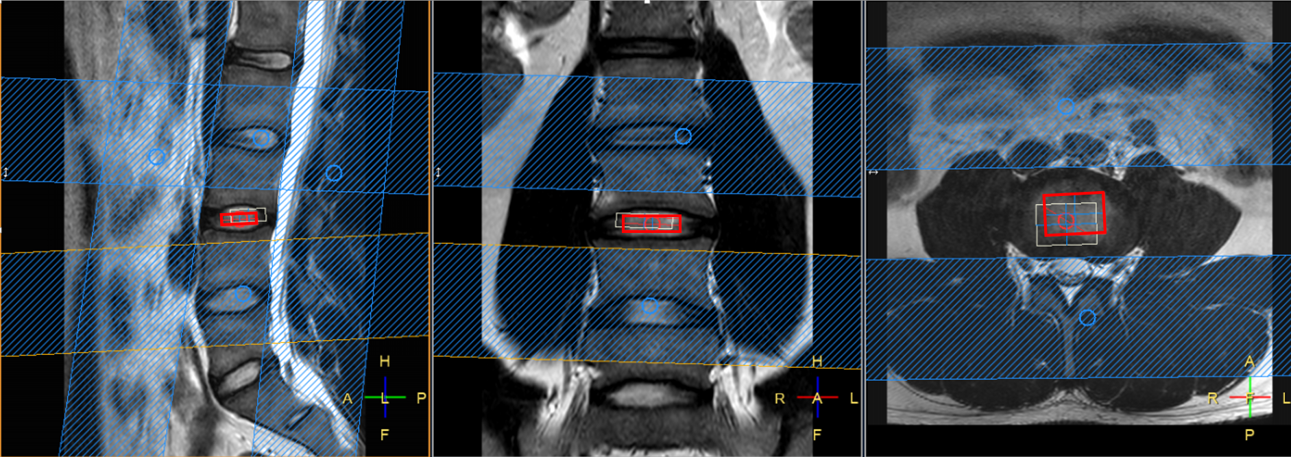
In this study, 9 discs (L3/L4, Pfirrmann grade=2-3) in 9 healthy volunteers (age=25-50years, 4 males) were examined on a 3T MRI scanner (Achieva, Philips). To assess the effect of breathing, phase images were acquired at two different states of the breathing cycle (expiration and inspiration) using a mid-sagittal slice over the lumbar spine (T1w-sequence, TE/TR=25/29ms, slice thickness=10mm, acquisition matrix=144x141).Disc MRS using Point RESolved Spectroscopy (single voxel (VOI) sequence, volume=17x6x25 mm, TE/TR=29/1800ms, 120 free-induction-decays) was performed during free-breathing. Iterative shimming of the magnetic field, as well as water suppression with variable pulse power and optimized relaxation delays (VAPOR) were used. To minimize signal contamination from tissue outside VOI, four saturation slabs were applied around the VOI (Fig.1).
The novel MRS-method was implemented on the Anaconda platform with the Python distribution (Vers.3, Anaconda Software Distribution). The postprocessing included truncation of free-induction-decays with a Tukey window and singular value decomposition to correct for phase effects due to respiratory effects while minimizing the noise. Optimal weights and phase shifts for each individual free-induction-decay were determined, searching for a solution to maximize the proteoglycan peak of the corresponding MRS-spectrum. The weighted spectra were then summed-up to one final spectrum.
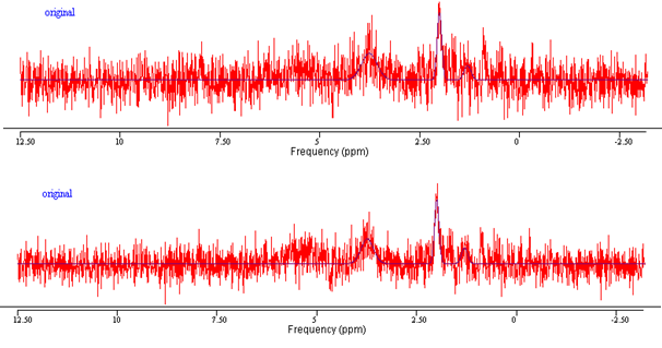
To display the effect of respiration, MRS-spectra were reconstructed both with and without correction of respiratory-induced phase effects.
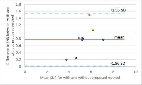
Lactate (1.33ppm) and proteoglycan peaks (2.04ppm), as well as metabolites associated with collagen breakdown (3.3-4ppm) were modelled in the final spectrum using the AMARES package4 in jMRUI (Vers.5.0)5. The SNR was calculated using jMRUI as the maximum of the model spectrum divided with the product of the standard deviation of the residue and the square root of the number of signal points in the free-induction-decay. To assess the improvement in SNR on a group level, Wilcoxon signed rank test was performed (Vers.27, SPSS Inc). The improvement in SNR with the proposed method was also shown in a Bland-Altman plot.
Results
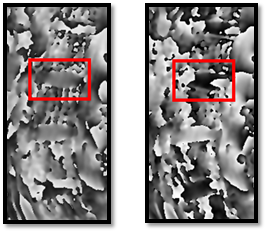
Successful MRS was performed on all individuals. The disc phase signal changed substantially between expiration and inspiration (Fig.2). There was significant difference (p=0.008) in SNR with and without the proposed method. The proposed postprocessing method gave for all spectra a higher SNR (mean gain=16%, range=5.5-30%), as shown graphically and numerically in (Fig.3-4).Discussion
This study is the first of its kind, using an automated postprocessing-method to enhance the quality of the molecular spectra of the discs. The method non-invasively generated spectra with higher SNR for all 9 volunteers with a maximum gain of almost 30%. This substantial improvement should be of great value for the otherwise low SNR measurement and might generate a more precise quantitation of the molecular content of the disc or shorten scan time. To verify the reliability of the method, a large cohort study, including also patients is encouraged.It is well known that respiratory motion can distort the MRS-measurement when performed in the thoracic region, but it may come as a surprise that this is also the case when measurements are performed far from that region, e.g. in the lumbar spine, here demonstrated by means of phase images. The phase effect can be explained by the change in magnetic field due to the relive difference in the lung volume between inspiration and expiration. Our novel automatic method adjusts the phase of each individual spectrum before merging them into the final spectrum, reducing the diminishing effect of breathing.
The recent evolution within computer-aided analysis have enabled mining of data to increase the diagnostic performance of MRS. In this study, a data-driven post processing method based on singular value decomposition modelling was used. As such, the method searches automatically for the solution that offer the highest SNR. Singular value decomposition was used as it has shown high feasibility for low SNR within other application areas6. Other methods or optimization of the present method might improve the quality of the MRS-spectrum further.
Conclusion
Present study shows that respiratory motion affects the phase signal in the disc and should be taken into consideration when evaluation the disc using MRS-measurements. The proposed method improved the quality of the MRS-spectrum and, thus, show feasibility in measuring the molecular disc content non-invasively during free-breathing. With an improved spectrum quality, smaller changes in the disc metabolite content can be detected, enabling longitudinal follow-up and comparisons between individuals.Acknowledgements
The authors would like to acknowledge the Swedish state under the agreement between the Swedish government and the county councils, the ALF-agreement(ALFGBG-792231, 813301, and 772931). We also want to acknowledge Nicolas Geades (Philips, Sweden) for clinical science expertise.References
1. Iatridis JC, MacLean JJ, O'Brien M, Stokes IA. Measurements of proteoglycan and water content distribution in human lumbar intervertebral discs. Spine (Phila Pa 1976). 2007;32(14):1493-7.
2. Keshari KR, Lotz JC, Link TM, Hu S, Majumdar S, Kurhanewicz J. Lactic acid and proteoglycans as metabolic markers for discogenic back pain. Spine (Phila Pa 1976). 2008;33(3):312-7.
3. Bolan PJ, Henry PG, Baker EH, Meisamy S, Garwood M. Measurement and correction of respiration-induced B0 variations in breast 1H MRS at 4 Tesla. Magn Reson Med. 2004;52(6):1239-45. 4. Vanhamme L, van den Boogaart A, Van Huffel S. Improved method for accurate and efficient quantification of MRS data with use of prior knowledge. J Magn Reson. 1997;129(1):35-43.
5. Naressi A, Couturier C, Devos JM, Janssen M, Mangeat C, de Beer R, et al. Java-based graphical user interface for the MRUI quantitation package. Magma. 2001;12(2-3):141-52.
6. Sung D, Risk BB, Owusu-Ansah M, Zhong X, Mao H, Fleischer CC. Optimized truncation to integrate multi-channel MRS data using rank-R singular value decomposition. NMR Biomed. 2020;33(7):e4297.
Figures