0250
Calibrationless B1 Mapping for Accurate Macromolecular Proton Fraction Mapping Using Relaxometry Constraints1University of Wisconsin-Madison, Madison, WI, United States
Synopsis
Macromolecular proton fraction (MPF) is an established myelin marker with confirmed clinical relevance, but with sensitivity to technological variations such as B1 field errors. We propose a method to derive B1 map for MPF correction from MPF data itself. The method is based on standard two-pool MT formalism enhanced with improved relaxometry constraints.
Introduction
Macromolecular proton fraction (MPF) is an established myelin marker with confirmed clinical relevance in a variety of neurological disorders. One drawback of MPF mapping is its substantial sensitivity to B1 field errors. While these can be measured by dedicated calibration protocols such as actual flip angle imaging (AFI)1, it comes at the expense of significant overhead (~3-4 min vs. ~7.5 min for MPF data collection)2. Recently, a possibility to estimate surrogate B1 map from MPF data itself was suggested3. The empirical observation of the global linear relationship between single-pool R1 and MPF in the absence of B1 errors was discovered. This allowed approximating B1 map with sufficient accuracy after learning calibration constants for a given acquisition protocol and scanner. In this work, we propose alternative treatment of the problem within fast MPF mapping framework, which is completely based on the two-pool MT model, thereby avoiding uncertainties associated with single pool approximations. We demonstrate that B1 estimation may be performed solely within the two-pool model formalism using normative estimates of relaxation constants.Theory
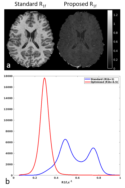
Our method is based on the recent findings that the use of underestimated values of longitudinal relaxation rate of macromolecular (bound) protons (R1b), a fixed parameter of the model, results in incomplete separation of macromolecular contributions from R1 of the free water protons (R1f), thereby inducing the macromolecular contrast in the R1f maps4. Proper modeling with more accurate estimates of R1b can enable the separation of macromolecular effects from R1f maps, which will make them consistent with the original model of this parameter (given by a sum of relaxation rates of a pure saline and contributions from paramagnetic substances5). The removal of macromolecular effects leads to spatially homogeneous R1f, as confirmed by results from our and other groups (Fig. 1). Here, we propose to use this fact for B1 map estimation within fast MPF mapping framework. Namely, instead of using B1 as a pre-measured map, we introduce it as a free parameter into the model while constraining R1f to its normative value.Methods
Data Acquisition: Experiments were performed on a 3.0T GE Discovery MR750 (Waukesha, WI) using an 8-channel head array in two healthy and three multiple sclerosis (MS) subjects after informed consent. The protocol included two SPGR and one MT-weighted SPGR measurement with optimized Z-spectra sampling and additional model constraints for fast 3-point MPF mapping as previously described6,7. Healthy volunteers were scanned with 1x1x2 mm resolution in 18 min; MS subjects were scanned with 2x2 GRAPPA-accelerated 1.3 mm-3 isotropic in 7.5 min8. Additionally, a B1 map was collected with AFI1 as a reference. Additional 3T MPF mapping dataset from different system (Philips 3T) was obtained from https://www.macromolecularmri.org/.Determination of Longitudinal Relaxation Constraints: We formulated our R1b estimation (submitted as a separate abstract to ISMRM 2021) using assumption that proper modeling interactions of free and bound protons with optimal R1b value should eliminate macromolecule-induced spatial variations from R1f, thereby minimizing the information content (as measured by histogram entropy function9). The fit yielded average value of R1b=6.5 s-1 across all subjects scanned on GE system. Mean R1f was 0.3045 s-1, which is consistent with previous studies3,4.
Processing: The MPF data were fit for proton density, MPF, and B1 error, while R1f and R1b were kept fixed to their normative values. As the resulting raw B1 map is defined only in brain tissue, its values in CSF and surrounding tissues were approximated using local polynomial fit smoothing10 weighted with MPF to measure a confidence of raw B1 estimation.
Results
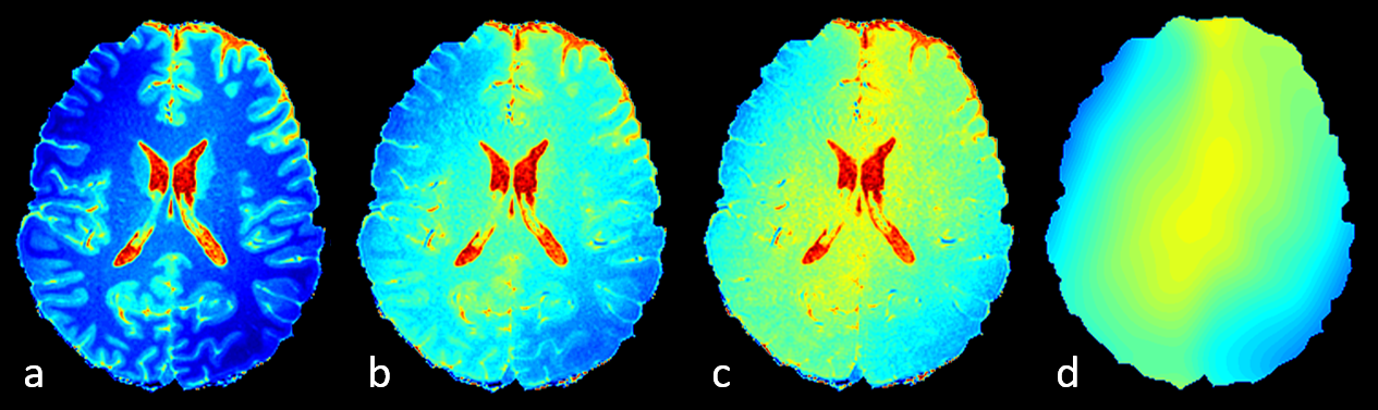
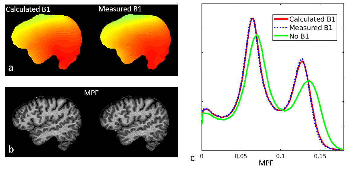
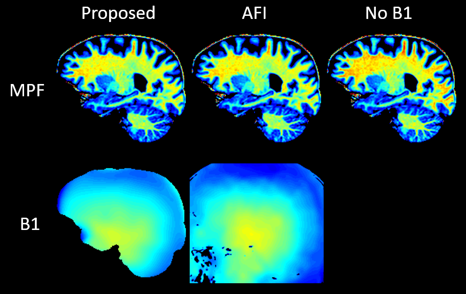
Figure 1 supports the use of R1f from improved two-pool modeling as a constrained parameter. Figure 2 illustrates the effect of improper selection of R1b (including previously proposed values for this parameter) on the calibrationless B1 field. Figure 3 presents results of applying our method for correction of MPF errors. Figure 4 demonstrates calibrationless B1 mapping on a different platform (Philips 3T), initially supporting the independence of the estimated relaxometry constants on the vendor.Discussion and Conclusions
We presented a calibrationless B1 mapping method, which derives from recent advances in two-pool MT modeling and is inspired by improved estimation of longitudinal relaxation rate of macromolecular protons. The method allows re-interpretation of the previous empirical B1 mapping method3 within two-pool model formalism, in which B1 mapping is performed as a standard two-pool model fit with constrained relaxometry constants. The method is directly applicable only in tissues that demonstrate MT effect. However, our results indicate that the B1 map may be reasonably well approximated within other non-MT areas such as CSF using standard assumption of B1 field smoothness. Another limitation is the potential sensitivity of calibrationless B1 maps to local deviations of R1f from its nominal value due to paramagnetic iron deposition in deep GM, which will be the subject of future studies.Acknowledgements
The work was supported by NIH (R01EB027087, R24NS104098) and GE Healthcare.References
1. Yarnykh VL. Actual flip-angle imaging in the pulsed steady state: a method for rapid three-dimensional mapping of the transmitted radiofrequency field. Magn Reson Med 2007;57(1):192-200. 2. Yarnykh VL. Time-efficient, high-resolution, whole brain three-dimensional macromolecular proton fraction mapping. Magn Reson Med 2016;75(5):2100-2106.
3. Yarnykh VL. Correction of B1 non-uniformity errors in fast macromolecular proton fraction and R1 mapping without B1 maps. In: Proc of ISMRM; 2020; Sydney, Australia. p 887.
4. Wang Y, van Gelderen P, de Zwart JA, Duyn JH. B0-field dependence of MRI T1 relaxation in human brain. Neuroimage 2020;213:116700.
5. Rooney WD, Johnson G, Li X, Cohen ER, Kim SG, Ugurbil K, Springer CS, Jr. Magnetic field and tissue dependencies of human brain longitudinal 1H2O relaxation in vivo. Magn Reson Med 2007;57(2):308-318.
6. Yarnykh VL. Fast macromolecular proton fraction mapping from a single off-resonance magnetization transfer measurement. Magn Reson Med 2012;68(1):166-178.
7. Samsonov AA, Mossahebi P, Anderson A, Velikina JV, Johnson KM, Johnson SC, Fleming JO, Field AS. High Resolution, Motion Corrected Mapping of Macromolecular Proton Fraction (MPF) In Clinically Acceptable Time Using 3D Undersampled Radials. In: Proc of ISMRM; 2014; Milan, Italy. p 3337.
8. Griswold MA, Jakob PM, Heidemann RM, Nittka M, Jellus V, Wang J, Kiefer B, Haase A. Generalized autocalibrating partially parallel acquisitions (GRAPPA). Magn Reson Med 2002;47(6):1202-1210.
9. Likar B, Viergever MA, Pernus F. Retrospective correction of MR intensity inhomogeneity by information minimization. Ieee Transactions on Medical Imaging 2001;20(12):1398-1410.
10. Mossahebi P, Yarnykh VL, Samsonov A. Analysis and correction of biases in cross-relaxation MRI due to biexponential longitudinal relaxation. Magn Reson Med 2014;71(2):830-838.
11. Nguyen TD, Spincemaille P, Gauthier SA, Wang Y. Rapid whole brain myelin water content mapping without an external water standard at 1.5T. Magn Reson Imaging 2017;39:82-88.
12. Zivadinov R, Reder AT, Filippi M, Minagar A, Stuve O, Lassmann H, Racke MK, Dwyer MG, Frohman EM, Khan O. Mechanisms of action of disease-modifying agents and brain volume changes in multiple sclerosis. Neurology 2008;71(2):136-144.
13. Khodanovich MY, Kisel AA, Akulov AE, Atochin DN, Kudabaeva MS, Glazacheva VY, Svetlik MV, Medvednikova YA, Mustafina LR, Yarnykh VL. Quantitative assessment of demyelination in ischemic stroke in vivo using macromolecular proton fraction mapping. Journal of cerebral blood flow and metabolism : official journal of the International Society of Cerebral Blood Flow and Metabolism 2018;38(5):919-931.
Figures