0208
Feasibility of Filter-exchange Imaging (FEXI) in Measuring Different Exchange Processes in Human Brain1Interdisciplinary Institute of Neuroscience and Technology (ZIINT), School of Medicine, Zhejiang University, Hangzhou, China, HangZhou, China, 2College of Biomedical Engineering and Instrument Science, Zhejiang University, Hangzhou, China, HangZhou, China, 3MR Collaboration, Siemens Healthcare, Shanghai, China, ShangHai, China, 4Department of Neurology, First Affiliated Hospital, School of Medicine, Zhejiang University, Hangzhou, China, HangZhou, China, 5Section on Quantitative Imaging and Tissue Sciences, NICHD, National Institutes of Health, Bethesda, MD, USA, Bethesda, MD, United States
Synopsis
In this study, we aim to explore the feasibility of Filter-exchange imaging (FEXI) in measuring different water exchange processes in human brain by modulating the diffusion filter (bf) and detection (b) blocks. We found the apparent exchange rate (AXR) estimated from a FEXI protocol with bf=250s/mm2 are significantly larger than those with bf=900s/mm2. Besides, the filter efficiency of FEXI with bf=250s/mm2 shows a strong correlation with vascular density estimated as the fraction of water exhibiting intravoxel incoherent motion (IVIM). Collectively, our current results demonstrate that FEXI targeting the vascular water could help characterize the intra-to-extravascular water exchange process.
Introduction
As a relatively new MRI technique, filter-exchange imaging (FEXI) targets the measurement of transmembrane water exchange with bf=800–900s/mm2 suppressing the fast diffusion component and measures the exchange processes between the two tissue water diffusion pools1-3. However, no studies have reported FEXI protocols to mainly filter out the blood water with limited effects on tissue water, which has the potential to measure the intra-to-extravascular water exchange process. In this study, we aim to explore whether FEXI could measure different water exchange processes via adjusting the filter and detection blocks. Two FEXI protocols were implemented on a 3T clinical MRI scanner with the first FEXI protocol targeting the intra-to-extravascular water exchange process and the second FEXI protocol following the available protocol2. Comparison between metrics derived from these two FEXI protocols and metrics from multi-b single PGSE measurement were performed on seven healthy volunteers in brain regional level.Methods
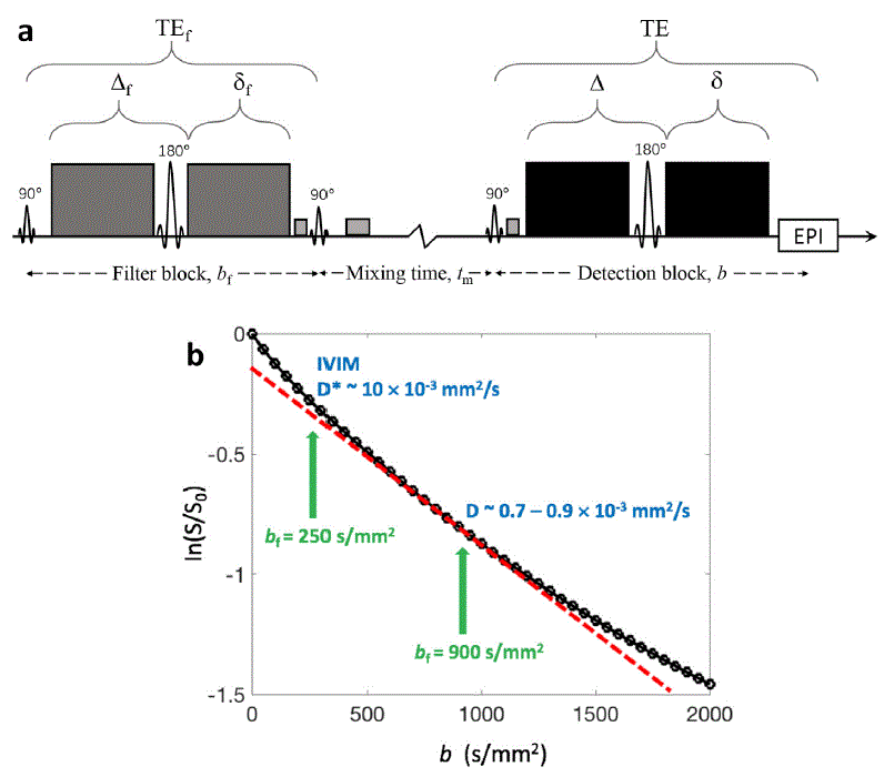
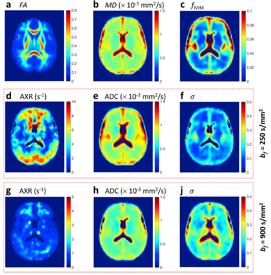
In this study, seven healthy subjects (age 24±2 years) were recruited and received MRI scans with a 3.0T MRI scanner (MAGNETOM Prisma, Siemens Healthcare, Erlangen, Germany). MRI scans included (1) 3D MP2RAGE T1-weighted images (1.0×1.0×1.2 mm3 resolution), (2) 3D SPACE T2-weighted images (0.9×0.9×1.0 mm3 resolution), (3) DTI with b=0s/mm2 (2 repetitions) , b=1000s/mm2 (20 directions), (4) IVIM DWIs acquired with a single PGSE: 15 b values from 0s/mm2 to 200s/mm2 with a step of 25s/mm2 and from 250s/mm2 to 500s/mm2 with a step of 50s/mm2, single acquisition at XY, YZ, and XZ direction for each b value, (5) first FEXI protocol with bf=250s/mm2 and b=0s/mm2 (3 repetitions), 250s/mm2 (6 repetitions), (6) second FEXI protocol with bf=900s/mm2, b=40s/mm2 (3 repetitions) and 900s/mm2 (6 repetitions). For both FEXI protocols, the directions of bf and b were always kept the same and acquired along three orthogonal directions (XY, YZ, and XZ), three : 25ms, 200ms, and 400ms were set. FEXI were also acquired with bf=0s/mm2 and shortest (25ms) for both protocols, as the equilibrium data for fitting. The FEXI sequence and two FEXI protocols are shown in Figure 1. All diffusion weighted images (DWIs) were acquired with 3.0×3.0 mm2 in plane resolution, slice thickness 5mm, 20 slices.Artifacts from eddy currents, motion and EPI distortion of DWIs were corrected with TORTOISE4. After pre-processing, the DTI data were fit to the non-linear DTI model in TORTOISE to calculate FA and MD. The IVIM data were fit according to,$$\frac{s_{b}}{s_{0}}=f_{IVIM}\exp(-bD^{*})+(1-f_{IVIM})\exp(-bD) [1]$$ where fIVIM, blood water fraction population; D*, apparent diffusivity of blood water consisting of both the blood water diffusivity and the pseudo-diffusion effect; and D, tissue water diffusivity. Details about this model can refer to previous studies5,6. For FEXI data, ADC'(tm) were computed from the two b (b1, b2) values in the detection block at each tm, $$ADC'(t_{m}) = -\frac{1}{b_{2}-b_{1}}\ln(\frac{s(t_{m},b_{2})}{s(t_{m},b_{1})}) [2]$$ Then, the ADC'(tm) at the three tm and bf=0 were fitted to $$ADC'(t_{m}) = ADC(1-\sigma\exp(-t_{m}AXR)) [3]$$ to obtain AXR, the filter efficiency (σ), equilibrium ADC.
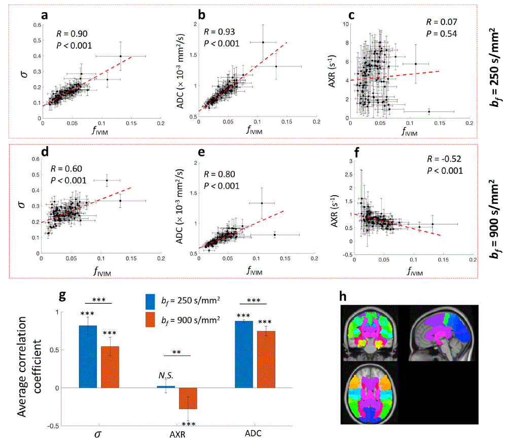
All DTI, IVIM and FEXI metrics were registered to the MNI152 space and classified into 121 different regions based on Jülich histological atlas7. Then Pearson correlation tests were performed among diffusion metrics of each region. For comparison of correlation coefficient (R), Fisher Z-transformation was performed on R at first, followed by Students’ t-test or paired Students’ t-test.
Results and Discussion
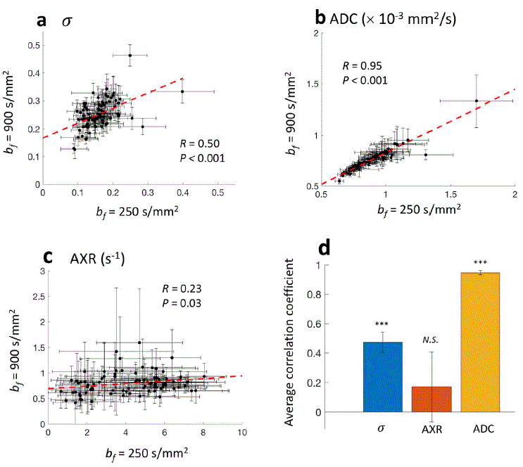
In figure 2, one slice of the standardized metric maps averaged from all the subjects are shown. Clear differences are observed between metrics of FEXI at bf=250s/mm2 and 900s/mm2. Figure 3 shows the results of correlation analysis between FEXI metrics and vascular density metric, fIVIM. The σ and ADC of FEXI at bf=250s/mm2 shows significant, almost linear correlation with fIVIM (Figure 3(a, b)). For FEXI at bf=900s/mm2, the averaged correlations between σ and ADC with fIVIM are significantly smaller than bf=250s/mm2 (P < 0.001) (Figure 3(g)). At the bf=250 s/mm2, the σ for the biexponential diffusion in brain tissue is expected to be low. Taking the diffusion data in human visual cortex8, the expected σ is only 0.06, whereas the expected σ are 0.29 for GM and 0.14 for WM if one uses the bicompartmental diffusion results from the IVIM data (Eq. (1)) in which the fast diffusion component is from vascular water. Taking these results together, we speculate that FEXI at bf=250s/mm2 measures water exchange between vascular and extravascular component. In Figure 4(c), no significant correlation is observed between the AXR of FEXI at bf=250s/mm2 and bf=900s/mm2, which further suggests the different water exchange processes between the two FEXI protocols. The results of FEXI at bf=900s/mm2 in this study are well consistent with the previous study2. Most literatures on AXR measured with FEXI at intermediate bf (~900 s/mm2) interpret it as intra- and extracellular water exchange rate1-3, 9-11, but more work is still needed to further understand the physiological basis of the apparent fast and slow diffusion components in brain tissue and the exchange between these two water diffusion pools.Conclusion
We have demonstrated the feasibility of FEXI in detecting different exchange processes in-vivo. FEXI at bf=250s/mm2 reveals the exchange between intravascular and extravascular water, whereas FEXI at bf =900s/mm2 measures exchange related to the bi-compartmental diffusion in brain tissue.Acknowledgements
No acknowledgement found.References
1. Lasič S, Nilsson M, Lätt J, et al. Apparent exchange rate mapping with diffusion MRI. Magnetic Resonance in Medicine 2011; 66: 356-365. DOI: 10.1002/mrm.22782.
2. Nilsson M, Latt J, van Westen D, et al. Noninvasive mapping of water diffusional exchange in the human brain using filter-exchange imaging. Magn Reson Med 2013; 69: 1573-1581. 2012/07/28. DOI: 10.1002/mrm.24395.
3. Lampinen B, Szczepankiewicz F, van Westen D, et al. Optimal experimental design for filter exchange imaging: Apparent exchange rate measurements in the healthy brain and in intracranial tumors. Magn Reson Med 2017; 77: 1104-1114. 2016/03/13. DOI: 10.1002/mrm.26195.
4. Pierpaoli C, Walker L, Irfanoglu M, et al. TORTOISE: an integrated software package for processing of diffusion MRI data. ISMRM 18th Annual Meeting 2010.
5. Iima M and Le Bihan D. Clinical Intravoxel Incoherent Motion and Diffusion MR Imaging: Past, Present, and Future. Radiology 2016; 278: 13-32. 2015/12/23. DOI: 10.1148/radiol.2015150244.
6. Le Bihan D. Magnetic resonance imaging of perfusion. Magn Reson Med 1990; 14: 283-292. 1990/05/01. DOI: 10.1002/mrm.1910140213.
7. Eickhoff SB, Stephan KE, Mohlberg H, et al. A new SPM toolbox for combining probabilistic cytoarchitectonic maps and functional imaging data. NeuroImage 2005; 25: 1325-1335. DOI: https://doi.org/10.1016/j.neuroimage.2004.12.034.
8. Le Bihan D. The 'wet mind': water and functional neuroimaging. Phys Med Biol 2007; 52: R57-90. 2007/03/22. DOI: 10.1088/0031-9155/52/7/R02.
9. Åslund I, Nowacka A, Nilsson M, et al. Filter-exchange PGSE NMR determination of cell membrane permeability. Journal of Magnetic Resonance 2009; 200: 291-295. DOI: https://doi.org/10.1016/j.jmr.2009.07.015.
10. Tian X, Li H, Jiang X, et al. Evaluation and comparison of diffusion MR methods for measuring apparent transcytolemmal water exchange rate constant. Journal of Magnetic Resonance 2017; 275: 29-37. DOI: https://doi.org/10.1016/j.jmr.2016.11.018.
11. Lasič S, Oredsson S, Partridge SC, et al. Apparent exchange rate for breast cancer characterization. 2016; 29: 631-639. DOI: https://doi.org/10.1002/nbm.3504.
Figures