0199
Positive emotional training with real-time functional MRI amygdala neurofeedback increased hippocampal volume for PTSD1Laureate Institute for Brain Research, Tulsa, OK, United States, 2Electrical and Computer Engineering, University of Oklahoma, Tulsa, OK, United States, 3Laureate Psychiatric Clinic and Hospital, Tulsa, OK, United States, 4Neuroscience Department, George Mason University, Fairfax, VA, United States, 5Department of Psychological Science, University of Arkansas, Fayetteville, AR, United States, 6Stephenson School of Biomedical Engineering, University of Oklahoma, Norman, OK, United States
Synopsis
While small hippocampal volume is a prevalent neurostructural abnormality in posttraumatic stress disorder (PTSD), whether hippocampal atrophy is a reversible alteration or a permanent trait is unclear. This study examined a volume change among hippocampal subfields due to positive emotion training with left amygdala (LA) fMRI neurofeedback (LA-NF) in PTSD participants. A significant volume increase was seen in the left CA1-head region after the training. This indicates that the small hippocampus in PTSD is a reversible alteration in a part of the subfields and that positive emotion training with LA-NF could induce a hippocampal volume recovery.
INTRODUCTION
Smaller hippocampal volume has been reported in posttraumatic stress disorder (PTSD) compared to both trauma-exposed controls without PTSD and non-exposed individuals. While an association between hippocampus dysfunction and PTSD deficits has been suggested, several studies indicated that small hippocampal volume may be a trait risk factor for PTSD but may not be associated with the disease state1-3. The trait- or state-dependent alteration could also differ among the hippocampal subfields. The present study examined the longitudinal hippocampal volume changes due to positive emotional training with left amygdala (LA) real-time fMRI neurofeedback (LA-NF) in combat veterans with PTSD.METHODS
Male combat veterans (21 to 48 years old) who met the DSM-IV-TR criteria for PTSD participated in the study. The participants were trained to increase the neurofeedback signal from the LA (experimental group [EG], N=20) or a brain region not involved in emotion processing (control group [CG], N=9) by recalling a positive autobiographical memory. The pre- and post-training structural MRI brain images were processed with FreeSurfer (v7.1.1) to evaluate the hippocampal subfield volumes. Volumes in 14 hippocampal subfields for each hemisphere (28 areas in total) were evaluated. Hippocampal volumes for healthy male controls (HC, N=43) were also examined to evaluate the baseline abnormality in PTSD. We used the longitudinal processing pipeline4, 5 for evaluating the volume change due to the rtfMRI-nf training in PTSD participants. The baseline hippocampal subfield alterations were examined by comparing the PTSD participants' pre-training volumes to the HC participant using linear model analysis with a diagnostic group (PTSD, HC) and covariates of age and the estimated total intracranial volume (eTIV). The percent volume change between the pre- and post-training scans was evaluated for PTSD participants and tested with linear model analysis with the condition (EG, CG) and covariates of age and the eTIV.RESULTS
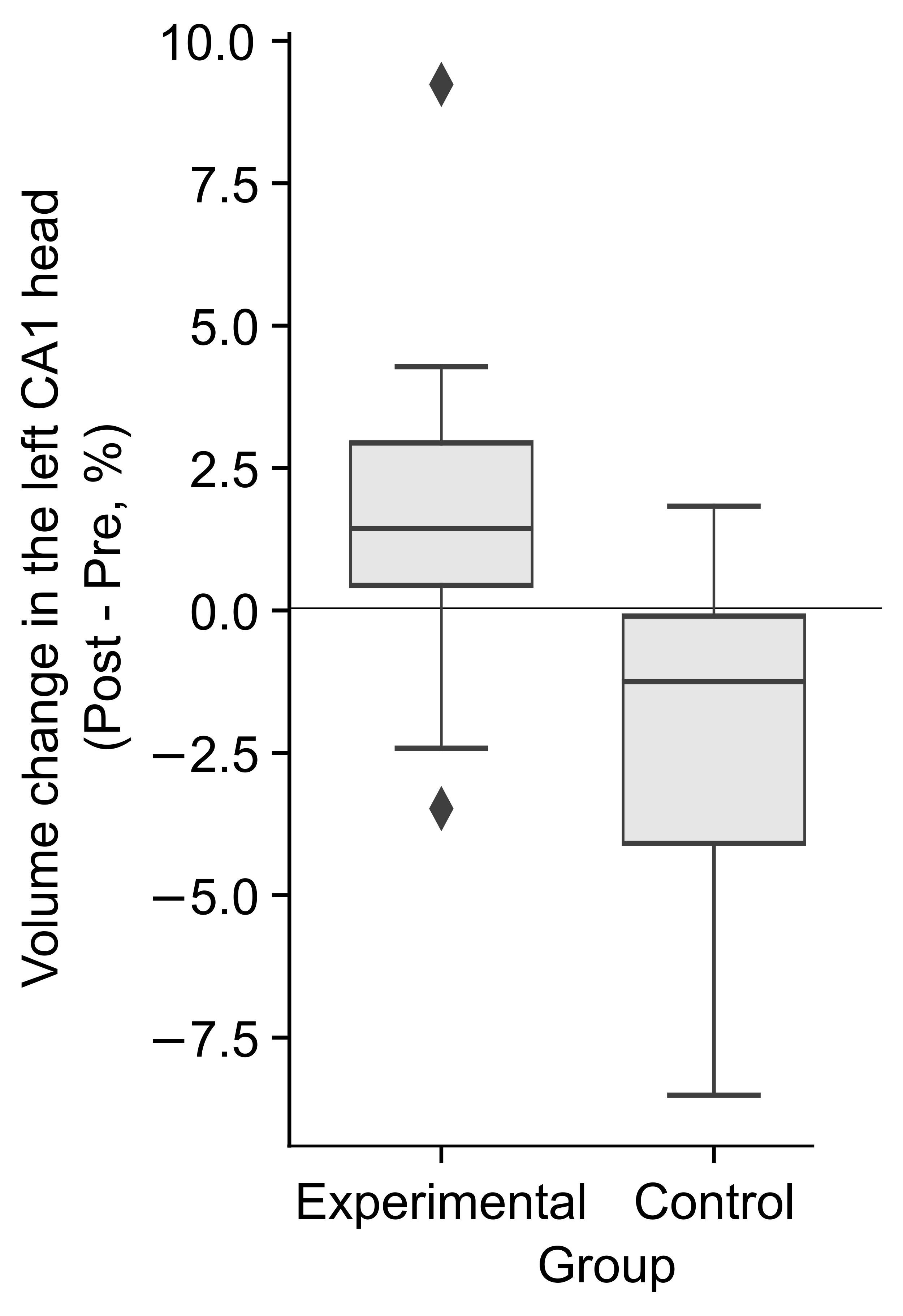
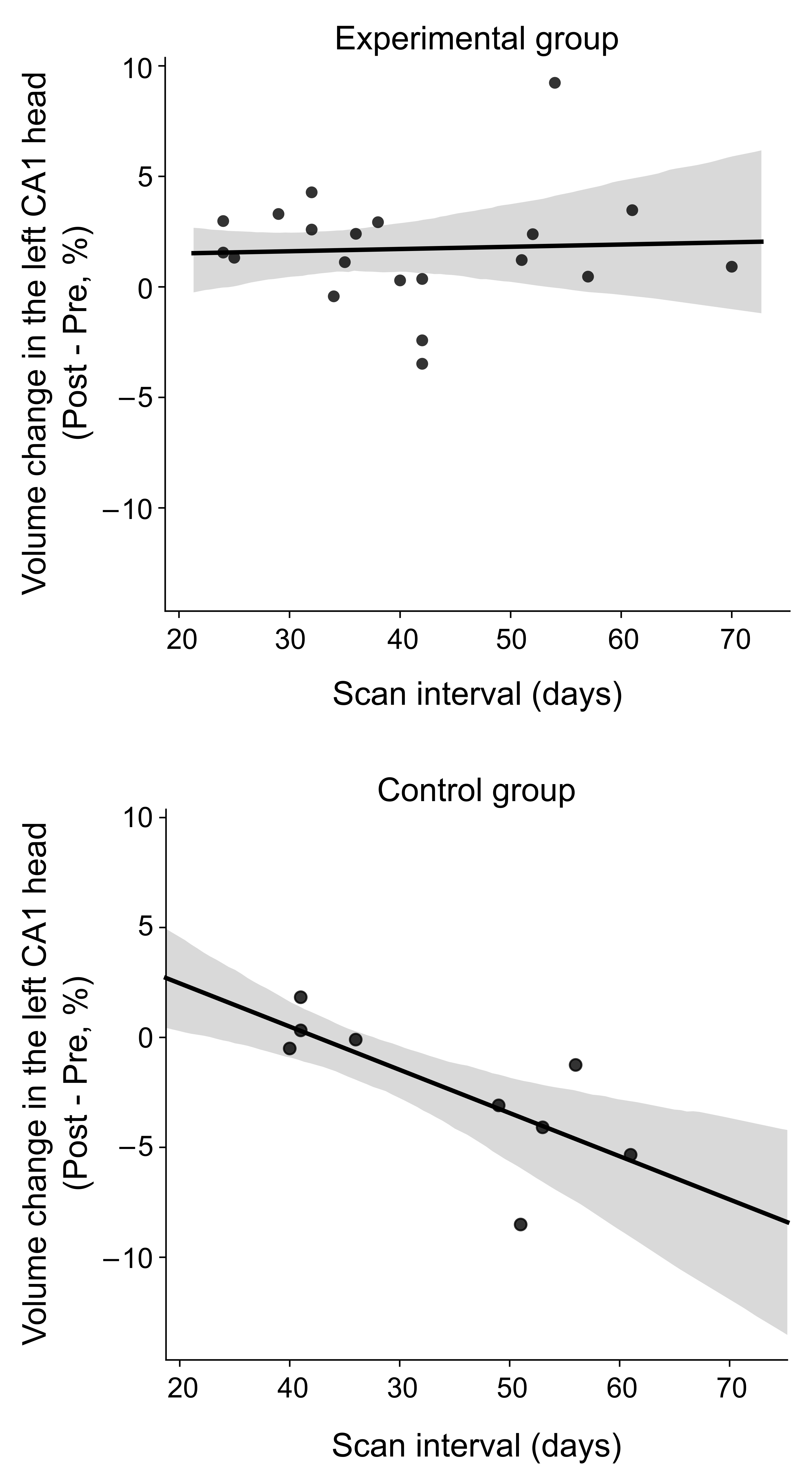
A significant group difference between the EG and CG was found in the percent volume change of the left CA1 head region (p<0.05 with false discovery rate correction). At the baseline, this region had the most significant volume reduction in PTSD compared to HC (d=-0.57, t=-2.35, p=0.022) but no significant difference was seen between the EG and CG in PTSD. The EG showed a significant volume increase (d=1.17, t=2.93, p=0.014) after the training (Fig. 1). In contrast, the CG showed a significant volume decrease (d=-1.07, t=-2.68, p=0.025) in this region and the volume change in CG was negatively correlated with interval days between the scans (d=-0.96, t=-2.31, p=0.059; Fig. 2).The EG showed significant symptom decrease measured with the Clinician-Administered PTSD Scale (CAPS) and Montgomery-Åsberg Depression Rating Scale (MADRS) after the sessions (CAPS, d=-1.87, t=-3.38, p=0.005; MADRS; d=-1.75, t=-3.81, p=0.001). Although the CG also showed a symptom decrease, the difference between the sessions was not significant (CAPS, d=-1.32, t=-1.75, p=0.124; MADRS, d=-0.99, t=-1.40, p=0.199). No significant association between the hippocampal subfield volume change and symptom change was found.
DISCUSSION
The hippocampal volume in the left CA1 head region increased after LA-NF positive emotion training for PTSD participants. Hippocampal plasticity and vulnerability to stress are variable between the subfields. Rodent studies demonstrated that the left CA1 is the most plastic and vulnerable region in the hippocampus6-8. The selective vulnerability of the left CA1 has been suggested for human adults too9. The CA1's association with autobiographical memory recall is also indicated10. These support the current finding of the selective volume recovery in the left CA1 head with the positive autobiographical memory recall training utilizing the LA-NF. The volume decreases for the left CA1 head in the control group had a trend of negative association with the scan interval, suggesting that the volume reduction could be ongoing in PTSD patients. This is also consistent with a study reporting a negative correlation between PTSD duration and hippocampal volume for combat veterans11.While no significant association between the volume increase and symptom recovery was found, this could be because the symptom scales could not measure the hippocampus functions, such as autobiographical memory recall, extinction memory recall, negatively-biased memory recall, and response inhibition. The deficit of these functions could worsen the symptom in the long-term, but the CAPS and MADRS would not be sensitive to measure the functional recovery associated with hippocampus volume change.
CONCLUSION
The small hippocampus in PTSD is not a permanent trait but a reversible alteration in a part of the subfields. Positive emotional training with left amygdala fMRI neurofeedback could induce a hippocampal volume recovery.Acknowledgements
This research was supported by W81XWH-12-1-0697 award from the U.S. Department of Defense, the Laureate Institute for Brain Research, and the William K. Warren Foundation. The data of non-trauma-exposed healthy males were provided by NIMH/NIH grant R01 MH098099. The authors report no biomedical financial interests or potential conflicts of interest.References
1. O'Doherty, D.C., K.M. Chitty, S. Saddiqui, et al., A systematic review and meta-analysis of magnetic resonance imaging measurement of structural volumes in posttraumatic stress disorder. Psychiatry Res, 2015;232(1):1-33.
2. Tischler, L., S.R. Brand, K. Stavitsky, et al., The relationship between hippocampal volume and declarative memory in a population of combat veterans with and without PTSD. Ann N Y Acad Sci, 2006;1071(1):405-9.
3. Gilbertson, M.W., M.E. Shenton, A. Ciszewski, et al., Smaller hippocampal volume predicts pathologic vulnerability to psychological trauma. Nat Neurosci, 2002;5(11):1242-7.
4. Reuter, M., N.J. Schmansky, H.D. Rosas, et al., Within-subject template estimation for unbiased longitudinal image analysis. Neuroimage, 2012;61(4):1402-18.
5. Iglesias, J.E., K. Van Leemput, J. Augustinack, et al., Bayesian longitudinal segmentation of hippocampal substructures in brain MRI using subject-specific atlases. Neuroimage, 2016;141:542-555.
6. Coultrap, S.J., K.M. Nixon, R.M. Alvestad, et al., Differential expression of NMDA receptor subunits and splice variants among the CA1, CA3 and dentate gyrus of the adult rat. Molecular Brain Research, 2005;135(1):104-111.
7. Kawakami, R., Y. Shinohara, Y. Kato, et al., Asymmetrical allocation of NMDA receptor epsilon2 subunits in hippocampal circuitry. Science, 2003;300(5621):990-4.
8. Shinohara, Y., H. Hirase, M. Watanabe, et al., Left-right asymmetry of the hippocampal synapses with differential subunit allocation of glutamate receptors. Proc Natl Acad Sci U S A, 2008;105(49):19498-503.
9. Bartsch, T., J. Dohring, S. Reuter, et al., Selective neuronal vulnerability of human hippocampal CA1 neurons: lesion evolution, temporal course, and pattern of hippocampal damage in diffusion-weighted MR imaging. J Cereb Blood Flow Metab, 2015;35(11):1836-45.
10. Bartsch, T., J. Dohring, A. Rohr, et al., CA1 neurons in the human hippocampus are critical for autobiographical memory, mental time travel, and autonoetic consciousness. Proc Natl Acad Sci U S A, 2011;108(42):17562-7.
11. Chao, L.L., K. Yaffe, K. Samuelson, et al., Hippocampal volume is inversely related to PTSD duration. Psychiatry Research: Neuroimaging, 2014;222(3):119-123.
Figures