0184
Novel Setup for 31P MRSI of the Human Tongue In Vivo at 7T1UMC Utrecht, Utrecht, Netherlands
Synopsis
Here we present the very first 31P MRS data in the human tongue at 7T. Three different receive coils were designed and evaluated to compare their performance: two each inside the mouth: a loop and also a saddle coil and a 3-channel loop array positioned externally. The transmit setup consists of an integrated 31P body birdcage coil for 31P transmit and an array of 8 1H dipoles for image registration. The loop coil seems to outperform the other two coils albeit that signals may originate from jaw. For voxels inside the tongue, relatively high phosphomonoesters were observed in all volunteers.
Introduction
Tongue cancer has a 5 year- survival rate of 50-60%. Current clinical practice is to use ultrasound image guided surgery to only remove the tumour without removing substantial parts of healthy tissue. Unfortunately, in many cases, the boundary of the excised specimen shows residual tumour tissue indicating extra surgery. An alternative to indicate tumour extent may be to reveal elevated cell proliferation by observing phospholipid metabolism1 with 31P MRSI. Here we present two intra-oral lingual – and one 3-channel external 31P Rx (receive) coils, combined with a distal 8 channel TxRx (transceiver) dipole array for 1H excitation placed inside a 31P full-body Tx birdcage coil integrated in a 7T MR system to investigate feasibility of non-invasive metabolic imaging of the tongue.Methods
Three versions of the 31P receive coils were constructed (Fig1). Version a) is a saddle coil formed around a mouth guard. Version b) is a simple loop coil to be inserted into the mouth such that it lies above the tongue with the head bent to maximize orthogonal orientation to the main field. Version c) is an array of three external loop coils which cover both cheeks as well as the mouth area.All coils had diode detuning implemented for detuning the receive coils during 31P transmit. This detuning circuitry was tested on the bench via a comparison between the transmission S-parameter measurements between the coil-under-test and a sniffer-loop. Additionally, the malfunctioning circuitry was verified before each RF pulse. For the phantom tests of coils b) and c) a spherical phantom containing two smaller spheres was placed inside the dipole array and the coil under test was positioned so as to be similarly located as the in vivo measurements. There are 4 different metabolites in the sphere: 200mM Pi in one smaller sphere; and 50mM each of PC, PE and GPC in the other smaller sphere.
For coil a), it was necessary to additionally submerge the coil in a 0.9% saline solution so as to provide sufficient load. The subjects were positioned head-first and supine with each of the coils used successively to perform the measurements. In each case, the possibility of coupling associated local SAR hotspots between the 1H TxRx array and the 31P receive coils was verified by a comparison of B1 maps – with and without the presence of 31P receive coils inside the 1H dipole array. A deviation of less than 10% was seen in all 3 cases, which was acceptable.
The protocol started with a localiser survey scan using the 1H array, followed by B0 shimming of the tongue area. An FID is acquired to determine the frequency offset in Hertz to place the metabolite of interest (PCr in vivo) at 0ppm. A 3D CSI in the transverse plane is then performed, with slightly varying parameters in vivo (FOV 224*224*225mm3 ; resolution 15*15*15mm3) than in the phantom (FOV 220*220*105mm3; resolution 22*22*7mm3) measurements. The CSI scans are performed at a low flip angle (12 degrees as Ernst angle) to accommodate a high bandwidth excitation at short TR; with a correspondingly short TE of 0.61ms and a TR of 60ms. RF block pulses are used giving a B1 of 4.5uT and a total scan time of 17 mins.
Post-processing of data was done in two main parts. First, the raw data were read into the Compass Framework (Tesla Dynamic Coils, Zaltbommel, the Netherlands), based on the WSVD channel combination method (for version c)). The PCr peak in vivo (Pi in the phantom) of this best voxel in the tongue area is then further processed to determine the area under the peak to account for B0 inhomogeneities. Similarly, the SNR for the best voxel is determined by the ratio of this area and the standard deviation of the spectral noise.
Results
Good background images could be obtained with the dipole array driven with a fixed amplitude and phase setting between the channels as optimized in a separate scan session (Fig 2). For all three receiver setups, good 31P MRSI could be obtained from the tongue, albeit that B0 shimming is severely compromised due to substantial air- tissue boundaries in the mouth (Fig 3). Apart from the data acquired with the loop, relatively high phosphomonesters (PME) are observed when compared to typically reported 31P MR spectra of muscle. In fact, the spectrum obtained from the loop may originate from the jaw muscles rather than the tongue, as judged from the background images and as it resembles the typically reported muscle spectra. The highest SNR of the voxel in the tongue area for volunteer 1 was 62 for the saddle, 174 for the loop and 96 for the external array.Discussion and Conclusion
Successful 31P MRSI can be obtained from the human tongue in vivo at 7T. Surprisingly, the external array outperformed the saddle coil, which may be due to B0 inhomogeneities and destructive phase coherence considering the relatively large voxels. Owing to the large chemical shift of 31P, despite suboptimal B0 shims, PME signals that are indicative of tumour grade can be well distinguished.
Acknowledgements
H2020-FETopen:NICIReferences
[1] K. Glunde, C. Jie, and Z. M. Bhujwalla, “Molecular Causes of the Aberrant Choline Phospholipid Metablism in Breast Cancer,” Cancer Res., vol. 64, pp. 4270–4276, 2004.Figures
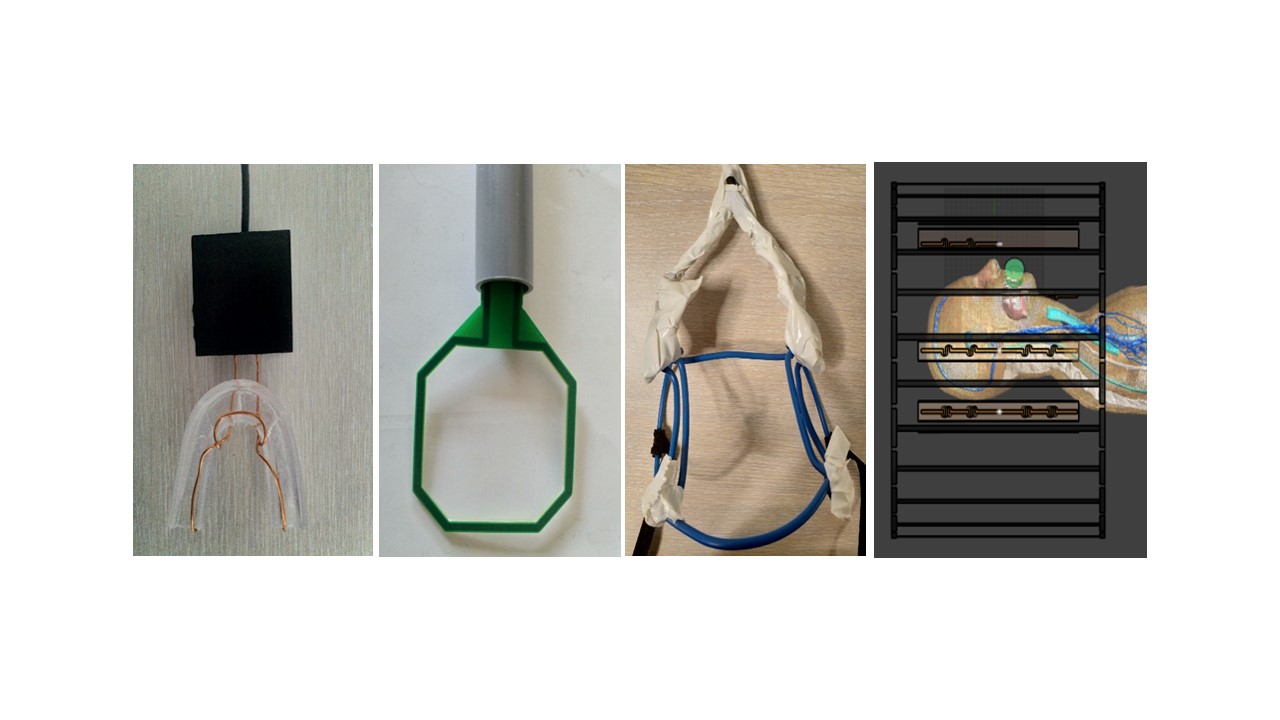
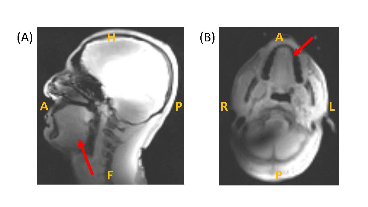
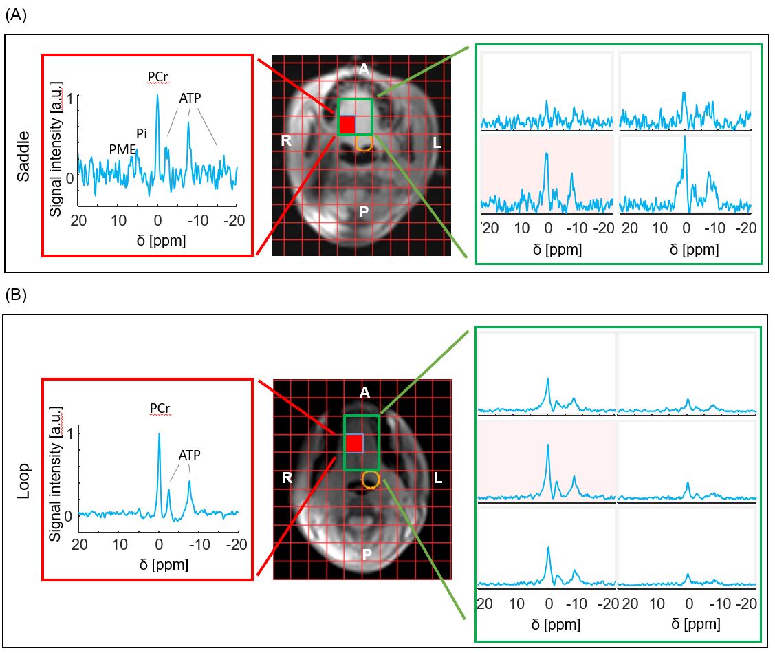
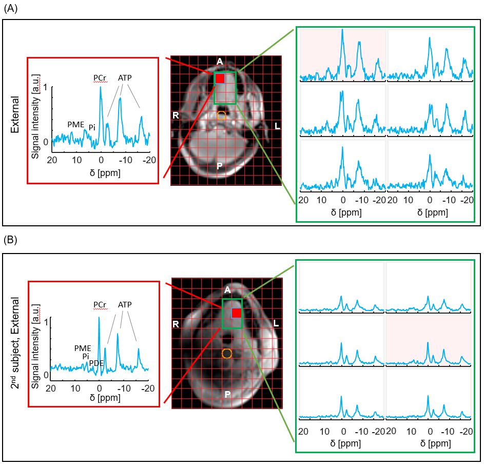
FIGURE 4: 31P MRSI results obtained from the external receiver coil in two subjects - also here PME is more intense, particularly in the first subject.