0179
A Self-decoupled 64 Channel Receive Array for Human Brain MRI at 10.5T1Center for Magnetic Resonance Research (CMRR), University of Minnesota Twin Cities, Minneapolis, MN, United States
Synopsis
Receive arrays play a major role in capturing the signal-to-noise ratio (SNR) and parallel imaging performance improvements at ultrahigh-field MRI. A novel self-decoupled 64-channel receive array was built for human brain imaging and compared to a 32-channel receive array at 10.5T/447MHz. Experiments demonstrated a maximum noise correlation of 0.31 in the 64-channel receiver. Experimental SNR comparisons showed 1.95 times more SNR averaged over the sample relative to the 32-channel array at 10.5T. The 10.5T 32 channel receiver was previously shown to have 1.81 times more average SNR gain over the industry standard 32 channel array at 7T.
Introduction
Signal-to-noise ratio (SNR) and parallel imaging (PI) advantages of ultra-high field MRI are beneficial to extensive clinical and research applications. Radiofrequency (RF) receive arrays are an integral requirement of realizing those advantages.In our previous work1,2, we compared a 10.5T 32-channel receiver (10.5T-32Rx) with a industry-standard (Nova Medical Inc. Wilmington, MA) 32-channel array operating at 7T and demonstrated the expected SNR and PI gains at the higher operating frequency of 447MHz. Here, we extend this work and present a novel 10.5T 64-channel receive array (10.5T-64Rx) and demonstrate further significant SNR gains compared to our previous receive array (10.5T-32rx).
Methods
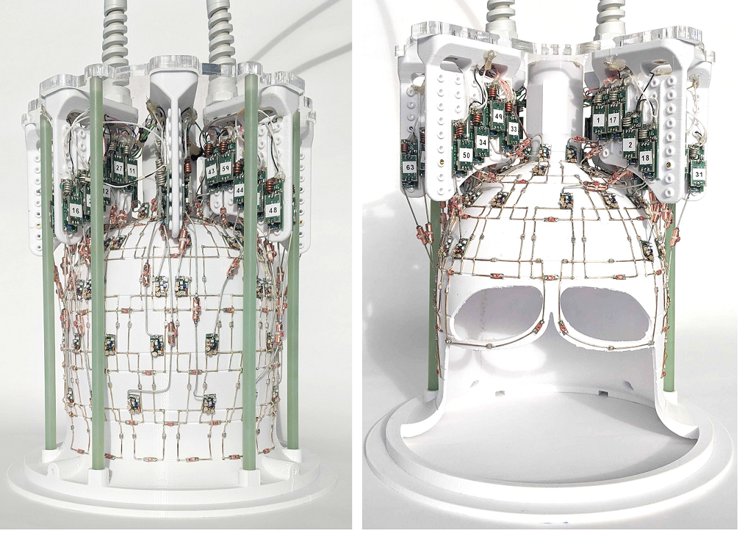
The 64 channel receive array was built using self-decoupling3 between azimuthally distributed elements of each row (there are six rows of loops). While there was overlap between rows along the z-direction (B0 direction), the elements within each row did not use overlap or inductive decoupling. Preamplifier decoupling2,4 was employed to further improve the receiver array noise correlation. Cable traps were incorporated to suppress shield-current-induced noise on preamplifier input coaxial cables. Figure 1 presents the as-built photograph of the 64-channel receive array.A 16-channel dual row loop transmitter was used in combination with the receive arrays5 (both the 64 channel and the 32 channel receivers can slide in and out of this 16 channel transmitter).
Identical protocols were used in both experimental measurements. Actual flip angle (AFI)6 maps were acquired and SNR acquisitions employed approximately fully relaxed (TR >> T1) transverse multi-slice GRE sequences. Details of our experimental protocol used to calculate SNR were published previously2. SNR calculations presented here were normalized for flip angle, voxel size, and bandwidth. Since the same MR scanner and phantom were used in both experiments, noise figure of the receive chain and T2* time constants were considered similar.
Results
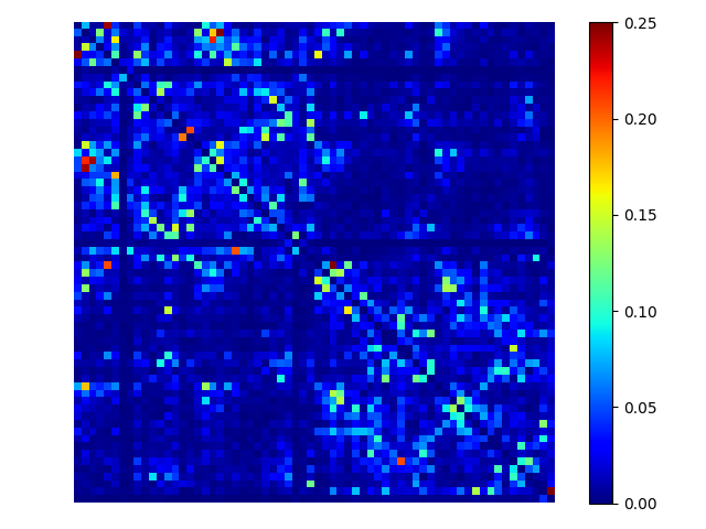
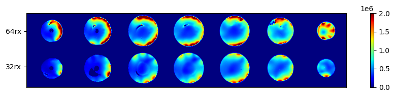
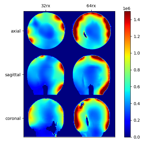
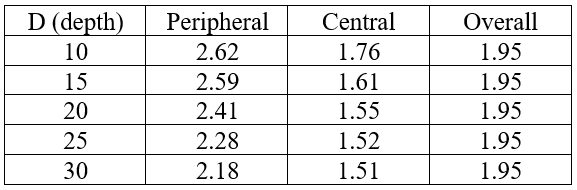
Figure 2 shows the experimental noise correlation measured at the scanner. Maximum (off-diagonal) noise correlation was 0.31. Figure 3 presents a comparison between the SNR of the 64-channel receiver and 32-channel receiver over axial slices. Figure 4 provides the SNR comparison in the central axial, sagittal and coronal slices of 3D SNR data sets. These figures demonstrate, qualitatively, the gain in SNR using the higher channel count receiver. Table 1 presents a quantitative comparison of SNR over the entire phantom. The 64-channel receiver provides 1.95 times more overall SNR on average, with 2.41x and 1.55x SNR gains at the peripheral and central ROIs (when the boundary between the peripheral and central ROIs is at 20mm from the phantom surface).Discussion
We present the successful design and construction of a 64-channel receive array for human brain imaging at 10.5T. The receive array takes advantage of preamplifier and self-decoupling principles and lays the groundwork for our future higher density receivers by mitigating the requirement of overlap or inductive decoupling. Whereas overlap/inductive receive array decoupling could be practically prohibitive for building 100+ channel receive arrays, our progress with self-decoupling receive arrays is an important development in that direction.There exist synergies between ultrahigh fields and high density receive arrays (see discussion in Ugurbil et al.7 and references therein). In agreement with this synergy, our results confirm previous expectations of SNR gains using higher density receivers at ultra-high fields. Previous simulations had indicated that using higher density of receive channels improves the SNR and would be especially advantageous in the peripheral regions of the brain (e.g. figure 3 of Keil and Wald8). Our results also demonstrate a more substantial SNR gain at the periphery versus the central ROIs. Parallel imaging analyses are also ongoing and will be presented.
Conclusion
We presented the first self-decoupled 64-channel receiver array for human head imaging, in this case operating at 10.5T, with SNR advantages demonstrated experimentally.Acknowledgements
The authors acknowledge NIH U01 EB025144, BTRC P41 EB027061, P30 NS076408, NIH S10 RR029672, and NIH R34AG055178 grants.References
1. Tavaf, N. et al. Developing High Channel Count Receive Arrays for Human Brain Imaging at 10.5T. Int. Soc. Magn. Reson. Med. (2020).
2. Tavaf, N. et al. A Self-Decoupled 32 Channel Receive Array for Human Brain Magnetic Resonance Imaging at 10.5T. (2020).
3. Yan, X., Gore, J. C. & Grissom, W. A. Self-decoupled radiofrequency coils for magnetic resonance imaging. Nat. Commun. 9, 3481 (2018).
4. Roemer, P. B., Edelstein, W. A., Hayes, C. E., Souza, S. P. & Mueller, O. M. The NMR phased array. Magn. Reson. Med. 16, 192–225 (1990).
5. Adriany, G. et al. Evaluation of a 16-Channel Transmitter for Head Imaging at 10.5T. in 2019 International Conference on Electromagnetics in Advanced Applications (ICEAA) 1171–1174 (IEEE, 2019). doi:10.1109/ICEAA.2019.8879131
6. Yarnykh, V. L. Actual flip-angle imaging in the pulsed steady state: A method for rapid three-dimensional mapping of the transmitted radiofrequency field. Magn. Reson. Med. 57, 192–200 (2007).
7. Ugurbil, K., Auerbach, E., Moeller, S., Grant, A., Wu, X., Van de Moortele, P.F., Olman, C., DelaBarre, L., Schillak, S., Radder, J., Lagore, R., Adriany, G., 2019. Brain imaging with improved acceleration and SNR at 7 Tesla obtained with 64-channel receive array. Magn Reson Med 82, 495-509.
8. Keil, B. & Wald, L. L. Massively parallel MRI detector arrays. J. Magn. Reson. 229, 75–89 (2013).
Figures