0178
A Quintuple-Tuned RF Coil for Whole Brain Multi-Nuclei Magnetic Resonance Imaging and Spectroscopy at 7T1Radiology, UMC Utrecht, Utrecht, Netherlands, 2Tesla Dynamic Coils, Zaltbommel, Netherlands, 3Biomedical Engineering, Eindhoven University of Technology, Eindhoven, Netherlands, 4Tesla Engineering Ltd, West Sussex, United Kingdom
Synopsis
Multi-nuclei magnetic resonance imaging and spectroscopy are interesting techniques to study metabolism, treatment efficacy tracking, and early diagnoses of many diseases. However, there are challenges to acquire high-quality MR images or spectra of nuclei other than proton, for example, much lower signal level, as well as the inconvenience of switching RF hardware in between scans, which leads to repositioning of the subject and extra preparation time. To overcome the above issues, we developed a quintuple-tuned RF coil for sensitive whole-brain scans, targeting five nuclei: 1H, 19F, 31P, 23Na and 13C. Bench tests and in-vivo scans have shown promising SNR.
Introduction
Metabolic imaging is powerful in the detection of treatment effects or for early diagnoses of many diseases. However, metabolic imaging via 1H MRSI is challenging primarily due to the dominant signals of water and lipids that can obscure the metabolite signals. When targeting other nuclei than 1H, metabolic imaging can be simpler, and sufficient sensitivity can be obtained at high fields. Yet due to the requirement of differently tuned coils, metabolic scans are normally not applied to patient studies that rely on conventional MRI as well. This abstract presents the design and performance of a quintuple-tuned head setup for an MRI system focused on merging anatomic with metabolic MRI (META-scan). Our setup combines a broadband eight-channel RF transceiver for 1H and 19F, as well as a triple-tuned local-receiver array for 31P, 23Na and 13C. Bench-top or scan measurements for all 5 nuclei are presented to indicate the efficiency. For sodium, phosphorus and proton, in vivo results from a human brain are shown.Methods
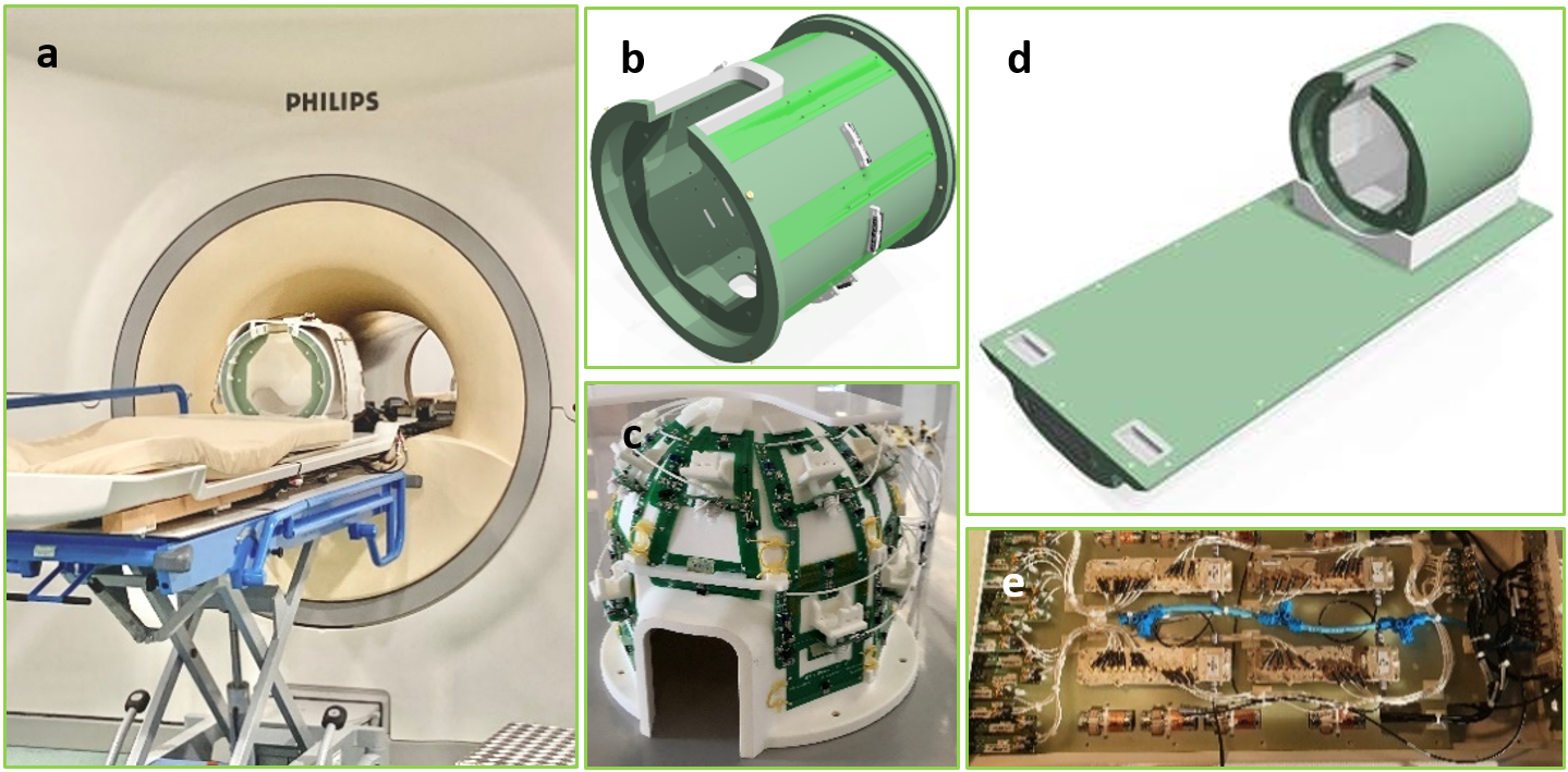
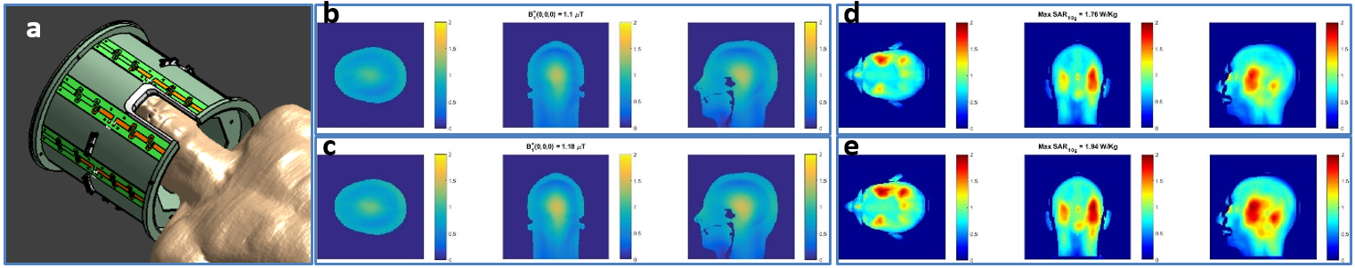
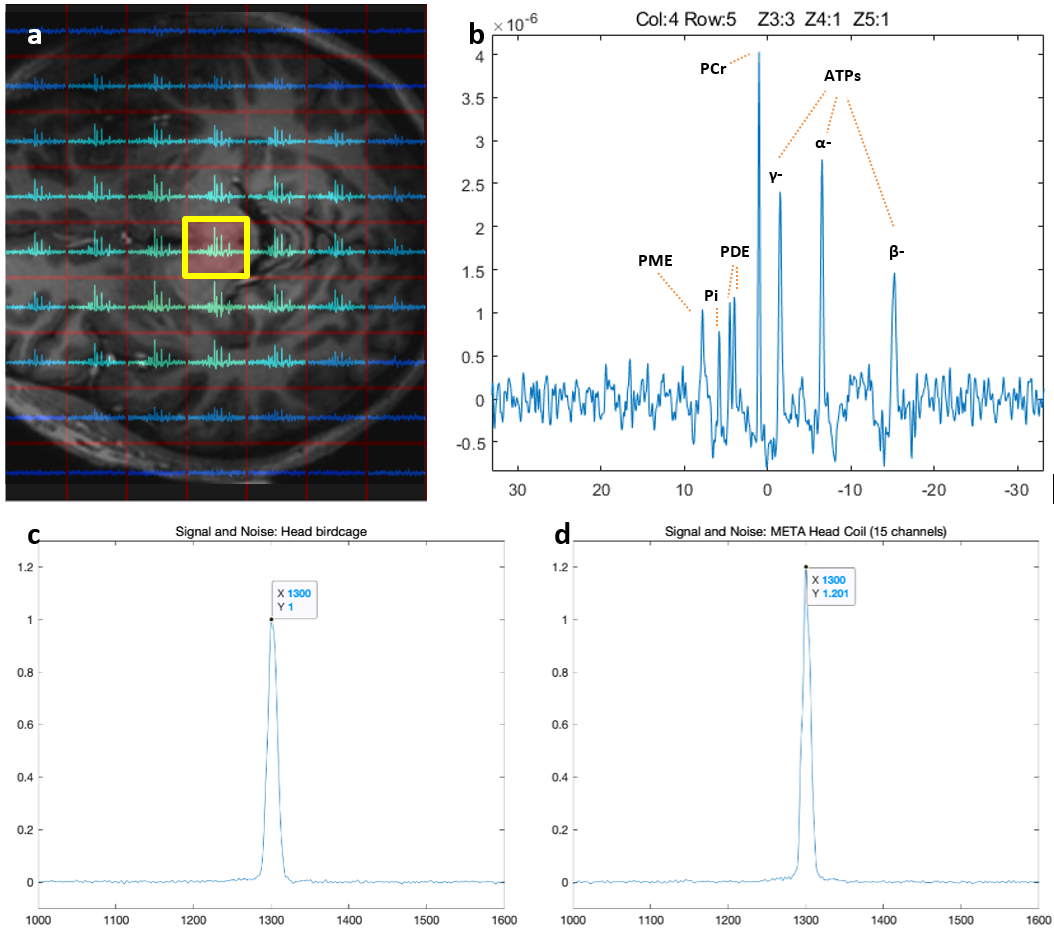
The META head coil (Figure 1a, 1d) contains an eight-channel dipole array (Figure 1b) as the RF transceiver for 1H and 19F, a fifteen-channel surface-loop array (Figure 1c) as RF receiver for 31P, 23Na and 13C, and a digital interface platform (Figure 1e) towards the scanner. The dipoles[1] are tuned at 290MHz, which is between the Larmor frequencies of 1H and 19F, taking advantage of the wide bandwidth of the dipole design. Each surface loop of the receiver array is dual-tuned for 31P and 23Na, and the third frequency is realized by switching an extra capacitance with a PIN diode to decrease the lower resonance from 79MHz(23Na) to 75MHz (13C). Each surface loop is directed to a low-loss diplexer to split signals of different frequencies. The split signals are fed to narrow-band preamplifiers and digital receivers, for 31P on one hand, 13C and 23Na on the other hand. The coil was interfaced to a 7T whole body MR system (Philips Healthcare, Best, NL). The system was equipped with an 8-channel Tx/Rx setup for 1H and 19F (8*2kW), a quadrature birdcage bore-coil[3] tuned for 31P, connected to a 25kW amplifier, and a Helmholtz clamp for 23Na excitation, connected to a 4 kW amplifier. In addition, the system was modified to accommodate multi-channel receiver array for all nuclei. B1+ and SAR simulations were performed using DUKE[4] (Sim4Life, Swiss) to assess safety and efficiency for 19F and 1H. Bench-top measurement of Q-factors of the receiver-loops in loaded and unloaded conditions were obtained and was compared to single-tuned loops to ensure high efficiency. 31P channel was also verified with a phantom scan comparison to a quadrature 31P birdcage[2]. An in vivo scan session was obtained with B1+ phase shimming for 1H, automated power adjustments, and B0 shimming, followed by a 3D TFE sequence of 6.5 minutes. In addition, a 3D sodium scan was obtained in 10 minutes with a TE of 1.35ms and TR of 90ms. Finally a 3D 31P CSI FID was obtained in 10 minutes using a flip angle of 13deg and TR of 71.1ms.Results
Proton and Fluorine: Figure 2 shows the simulation model, the simulated B1+ field and the local SAR at both 19F and 1H Larmor frequencies at 7T, with 1 Watt accepted power per channel, in quadrature mode. It indicates that, with the same amount of accepted power, the transmit fields for 1H and 19F are highly similar. Figure 3 shows the TFE images. 18µT was achieved at the center of the ROI, with 922 Watt forward peak power per channel.Phosphorus: The SNR in the center of META head coil is 11% higher compared to a highly efficient dual-tuned (31P and 1H) head-birdcage[2] under identical scan conditions (i.e. same flip angle, TR, and load). Figure 4c-d show the signal-level comparison. Figure 4a-b show a slice of the 3D CSI, indicating good coverage over the entire brain and high SNR.
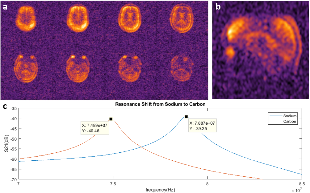
Sodium: Figure 5a-b show 23Na images of an in-vivo scan. Considering the very short T2* of 23Na, the used echo time (1.35ms) is far from optimal.
Carbon: Bench measurements show that the receiving performance of the 13C elements is similar to that of 23Na (at max 1dB loss): S21 between two pick-up probes next to an unloaded surface-loop shows that the Q-factor of the unloaded surface-loop does not change when shifting from the resonance of 23Na to that of 13C (Figure 5c).
Discussion and Conclusion
We have demonstrated a successful integration of an RF setup that could detect 23Na, 13C, 31P, 19F and 1H in a single scan session. Since tracers are generally required for 13C and 19F, we show natural-abundance 3D images of only 31P, 23Na and 1H, in a healthy human brain. Since the frequency of 13C is close to 23Na and 19F close to 1H, we do not expect the sensitivity to be different for these nuclei. Excluding the preparation time, the acquisition time for the 1H, 31P and 23Na scans can be less than 25 minutes in total, paving the way to incorporate metabolic MRI of all five nuclei in one scan session of acceptable scan time.Acknowledgements
I would like to thank Dutch Research Council (NWO) for funding the META-scan project.
References
[1] Raaijmakers, A.J., Italiaander, M., Voogt, I.J., Luijten, P.R., Hoogduin, J.M., Klomp, D.W. and van den Berg, C.A. (2016), The fractionated dipole antenna: A new antenna for body imaging at 7 Tesla. Magn. Reson. Med., 75: 1366-1374. https://doi.org/10.1002/mrm.25596
[2] Hendriks, AD, van der Kemp, WJM, Luijten, PR, Petridou, N, Klomp, DWJ. SNR optimized 31P functional MRS to detect mitochondrial and extracellular pH change during visual stimulation. NMR in Biomedicine. 2019; 32:e4137. https://doi.org/10.1002/nbm.4137
[3] van Houtum, Q, Welting, D, Gosselink, WJM, Klomp, DWJ, Arteaga de Castro, CS, van der Kemp, WJM. Low SAR 31P (multi‐echo) spectroscopic imaging using an integrated whole‐body transmit coil at 7T. NMR in Biomedicine. 2019; 32:e4178. https://doi.org/10.1002/nbm.4178
[4] Christ A, Kainz W, Hahn EG, et al. The Virtual Family—development of surface‐based anatomical models of two adults and two children for dosimetric simulations. Phys Med Biol. 2010;55:N23–N38.
[5] Froeling M: QMRTools: a Mathematica toolbox for quantitative MRI analysis. J Open Source Softw 2019; 4:1204.
Figures