0156
Mapping functional connectivity of thalamus subdivisions in obsessive-compulsive disorder1Huaxi MR Research Center (HMRRC), Functional and molecular imaging Key Laboratory of Sichuan Province, Department of Radiology, West China Hospital, Sichuan University, Chengdu, China, 2Sichuan University, Chengdu, China
Synopsis
Using connectivity-based parcellation technique, we segmented thalamus into two distinct subdivisions comprising superior thalamus and inferior thalamus based on their similarity in resting-state functional connectivity properties. We then compared the functional connectivity profiles of each thalamic subdivisions between the obsessive-compulsive disorder (OCD) patients and healthy control (HC), and revealed the disturbances of the superior/inferior thalamo-cortical and inferior thalamo-cerebellar circuitry in OCD patients. These findings suggested that thalamus subdivisions play different role in motor, cognitive, affective processes in OCD, which may underlie the pathophysiology of the disorder.
Introduction
The pathophysiology of obsessive-compulsive disorder (OCD) has been widely conceptualized within the cortico-striato-thalamo-cortical model 1, emphasizing the thalamic abnormalities implicated in OCD. Most previous neuroimaging studies investigating thalamic functional features in OCD have used the whole thalamus as the region of interest. However, the composition of thalamus is complex; neuroimaging data typically averaging over all voxels of the thalamus to provide a summary characterization at the cost of potentially missing important information of variation in connectivity across the topography of the thalamus may be damaging to network estimation. A recently proposed approach of connectivity-based parcellation (CBP) makes it possible to segment brain regions in vivo according to the functional connectivity properties and provide a good condensed representation of voxel-wise data for subsequent analyses, with resulting clusters being more homogeneous in terms of resting-state signal than, for example, cytoarchitectonic areas 2. The current study used mapping of internal thalamus subsegment derived from CBP approach to delineate precise neural circuitry alterations in patients with OCD.Materials & Methods
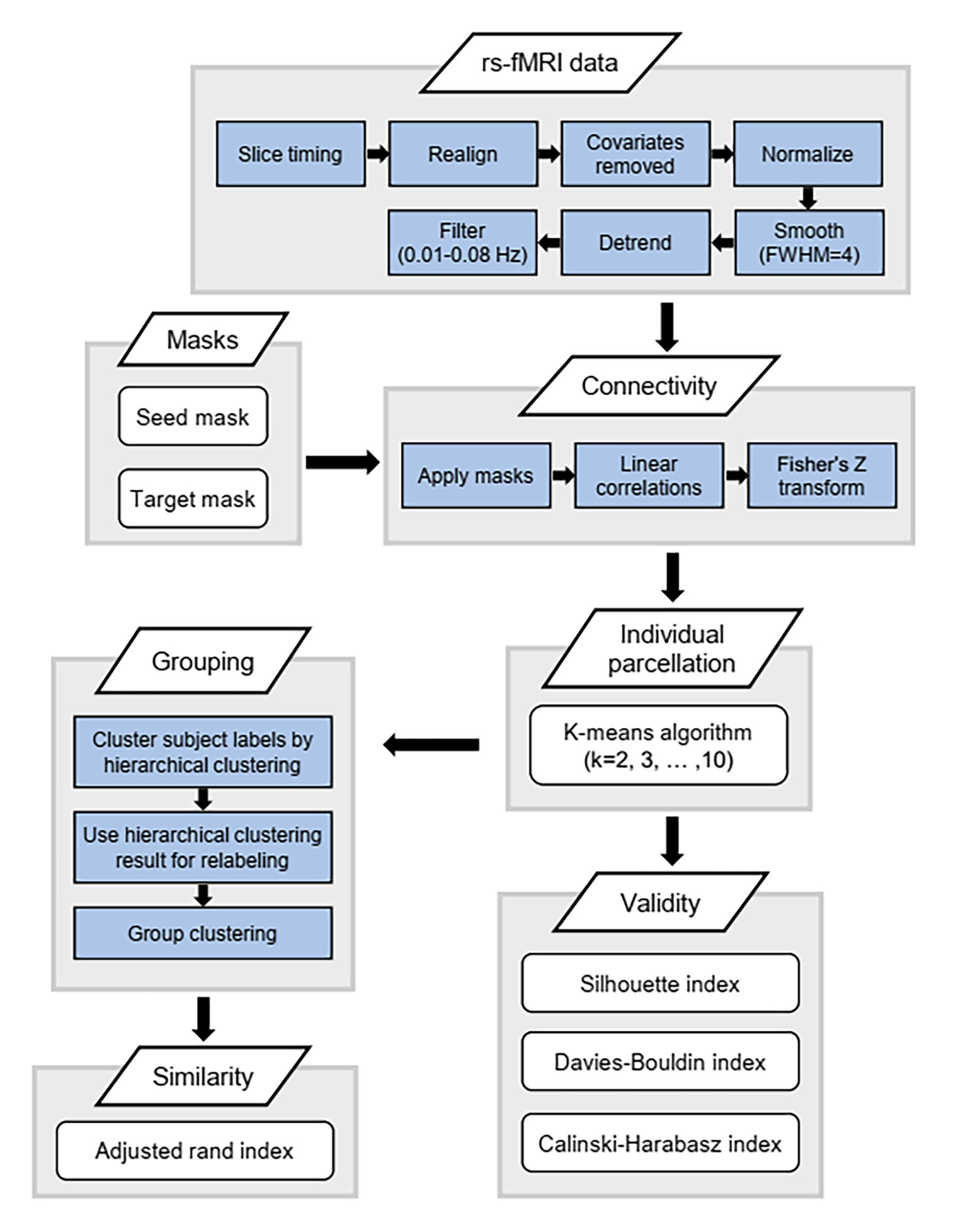
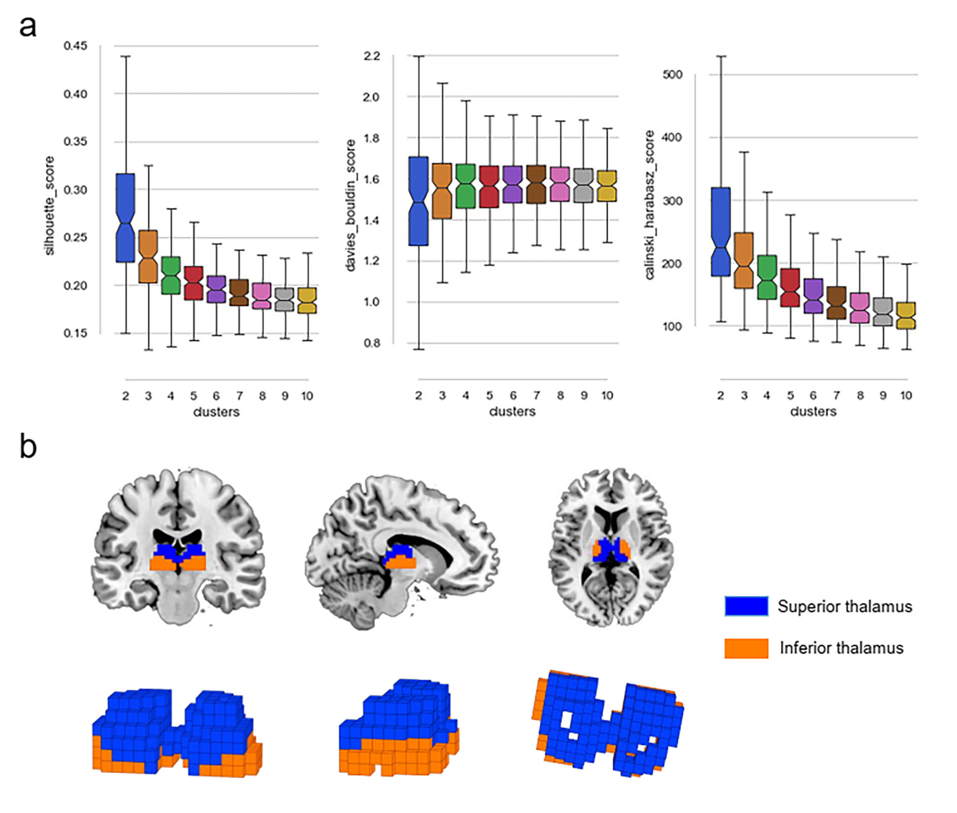
A total of 87 DSM-Ⅳ criteria diagnosed OCD patients and 90 age- and sex-matched healthy controls (HC) participated in this study after giving written informed consent. Among the patients, 73 individuals have never received medication treatment, and the remaining patients were medication-free for a minimum of 4 weeks prior to the scan. All participants were scanned using 3-Telsa GE magnetic resonance imaging (MRI) to acquire resting-state functional MRI data and high-resolution structural MRI data.Preprocessing of the resting-state functional images includes slice timing, realignment, regressing out nuisance signals, spatial normalization, smooth, detrend and filtering. Connectivity-based parcellation was conducted by using CBPtools 3 to segment the entire thalamus (both left and right parts together) into distinct subdivisions based on their resting-state functional connectivity patterns with the whole brain (Figure 1). Briefly, functional connectivity between each thalamic voxel and every voxel of the whole brain was calculated for each participant. Then, a k-means clustering was applied to the connectivity matrix to assign each thalamic voxel to a cluster, effectively grouping similar voxels based on their connectivity profiles and obtaining individual thalamic parcellations. To determine a group parcellation that best describes all included subjects, the individual clusterings were relabeled and the mode of the relabeled subject-wise clustering was computed and used as the group-level clustering. Several cluster quality metrics including the Silhouette Index, the Calinski–Harabasz index, and the Davies–Bouldin index, were obtained to find the appropriate number of clusters.
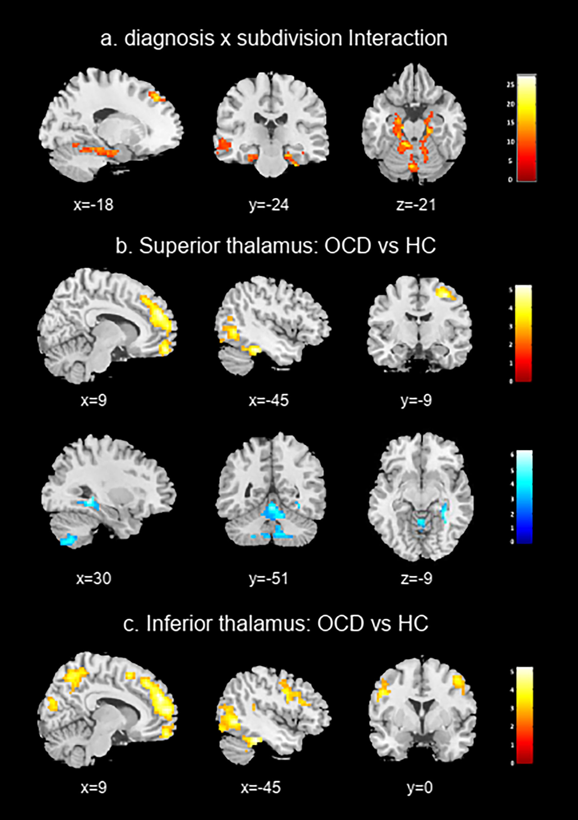
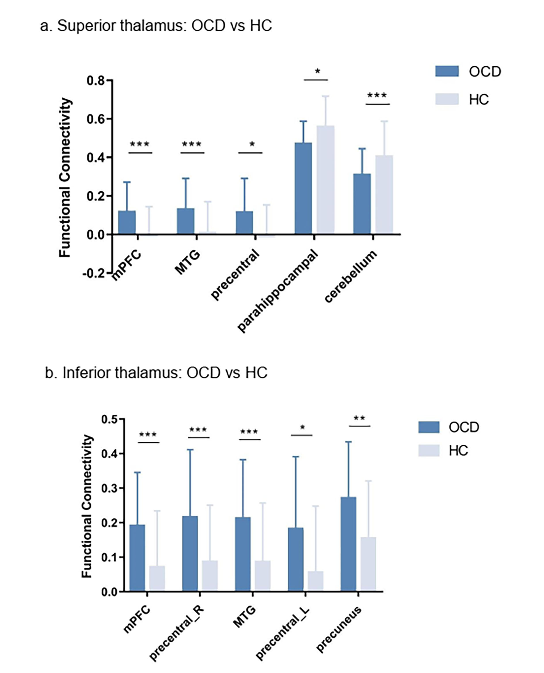
Finally, a full factorial analyses of variance was used to test the interaction between diagnosis and subdivision. To characterize different functional connectivity patterns of thalamic subdivisions between OCD patients and HC, voxel-based comparisons of functional connectivity maps of the thalamic subdivisions were performed. The significance threshold was set to p<0.005 at the voxel level, and FWE correction at the cluster level to p<0.05.
Results
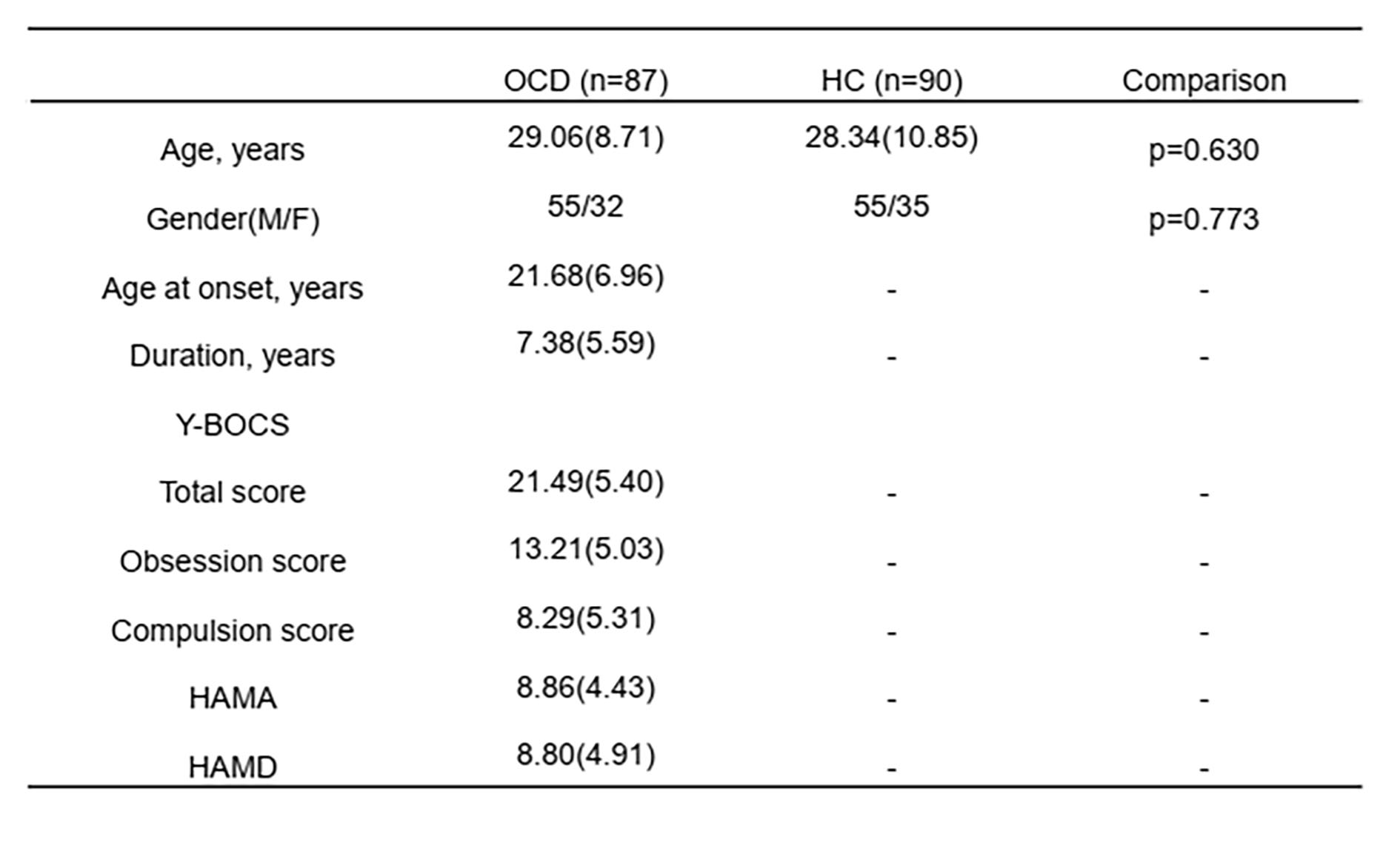
The demographic information and clinical characteristics of the participants are shown in Table 1.The Silhouette index, Davies–Bouldin index, and Calinski–Harabasz index all indicated the two-cluster separation into a superior and inferior subdivision fitted the input data best (Figure 2).
Full factorial analyses of variance revealed significant interactions between diagnosis and subdivision in several brain regions including the superior frontal gyrus, middle temporal gyrus, parahippocampal gyrus, and cerebellum. Relative to HC, OCD patients exhibited increased functional connectivity between the superior/inferior thalamus and the medial prefrontal cortex, middle/inferior temporal gyrus/fusiform and precentral gyrus. Additionally, functional connectivity between the inferior thalamus and the precuneus/cuneus was significantly increased in OCD patients. In contrast, the superior thalamus showed significantly decreased connectivity with the parahippocampal gyrus and cerebellum (Figure 3 & Figure 4).
Discussion & Conclusion
Using connectivity-based parcellation, the current study suggest that the thalamus is best represented with a bipartite superior-inferior subdivision. More importantly, distinct functional connectivity profiles of thalamic subdivisions between the OCD patients and HC were identified. Specifically. both superior and inferior thalamic subdivisions showed stronger connectivity with the regions of prefrontal cortex, temporal lobe and motor cortex in OCD patients, which may result from derailing the normal development of thalamic-cortical connectivity during the transition from adolescence to adulthood 4. Additionally, inferior thalamic subdivision exhibited stronger connectivity with the regions of posterior parietal and occipital cortex in OCD patients, which may be associated with abnormal visual processing and spatial attention. In contrast, weaker functional connectivity between the superior thalamic subdivision and the cerebellum may perhaps provide a neuroimaging evidence of imbalance of the cerebellar-thalamic inhibitory output in OCD patients. These findings establish differential abnormalities of thalamic subregional connectivity in OCD patients, which may underlie the neural mechanisms of the disorder.Acknowledgements
This study was supported by National Nature Science Foundation (Grant NO. 81671669), Science and Technology Project of Sichuan Province (Grant NO. 2017JQ0001).References
1. Stein DJ, Costa DLC, Lochner C, et al. Obsessive-compulsive disorder. Nature reviews Disease primers 2019; 5(1): 52.
2. Eickhoff SB, Yeo BTT, Genon S. Imaging-based parcellations of the human brain. Nature reviews Neuroscience 2018; 19(11): 672-86.
3. Reuter N, Genon S, Kharabian Masouleh S, et al. CBPtools: a Python package for regional connectivity-based parcellation. Brain structure & function 2020; 225(4): 1261-75.
4. Fair DA, Bathula D, Mills KL, et al. Maturing thalamocortical functional connectivity across development. Frontiers in systems neuroscience 2010; 4: 10.
Figures