0132
Wearable knee receive array coil for imaging at different flexion angles1Department of Electrical & Computer Engineering, New York University Tandon School of Engineering, Brooklyn, NY, United States, 2Bernard and Irene Schwartz Center for Biomedical Imaging, Department of Radiology, New York University Grossman School of Medicine, New York, NY, United States, 3Center for Advanced Imaging Innovation and Research, Department of Radiology, New York University Grossman School of Medicine, New York, NY, United States
Synopsis
Weight-bearing and kinetic MRI are important for measuring tibialfemoral joint dynamics, but are difficult to carry out using rigid knee coils that are typically designed to restrict, rather than enable, flexion motion. We explored off-the-shelf components and constructed a six-channel flexible knee coil with an elastic shell to maintain critical geometric overlap between neighbor coils. The array enables MRI during knee flexion while providing similar SNR compared to a state-of-the-art rigid commercial coil. We anticipate that the coil will be useful for weight-bearing or kinetic knee imaging in which rigid coils provide suboptimal SNR and/or severely restrict the desired posture.
Introduction
Musculoskeletal MRI can characterize articular and patellar cartilage and help detect degradation related to osteoarthritis. While clinical knee MRI is performed under static conditions, recent research has shown that weight-bearing and kinetic imaging provides new information on ligament and meniscus stress, and post-load recovery dynamics 1, 2, 3, 4. Such experiments are difficult to perform or are compromised by rigid knee coils that are typically designed to restrict, rather than enable, flexion motion. Several groups have used various strategies to develop flexible coils to accommodate flexion motion or improve anatomical conformability 5, 6, 7, 8, 9. To avoid increased coil conductor loss that can be associated with flexible substrates while also maintaining ease-of-assembly, we fabricated a flexible array out of coaxial cable-based loops. The loops were installed into a stretchable fabric shell with integrated pockets to maintain critical geometric decoupling during knee flexion. To explore the design space, we compared quality factor (Q) and SNR measurements from loops made with different coaxial cables and coil topologies, as well as with mechanical deformation that would occur during knee flexion. We used insights from the initial exploration to guide the development of a flexible six-channel array that we applied to knee imaging at different flexion angles.Methods
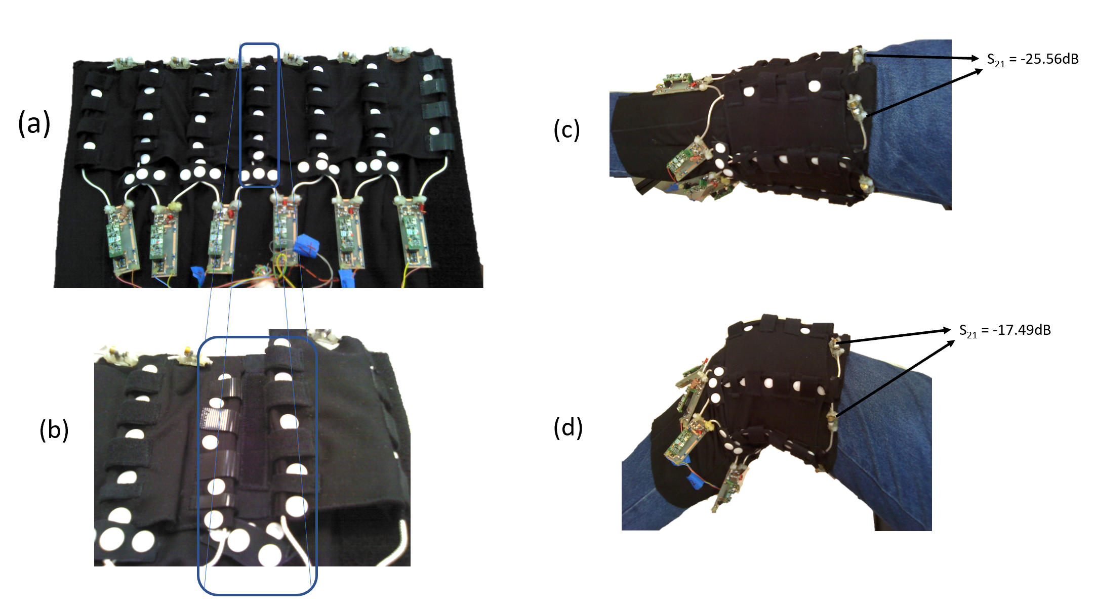
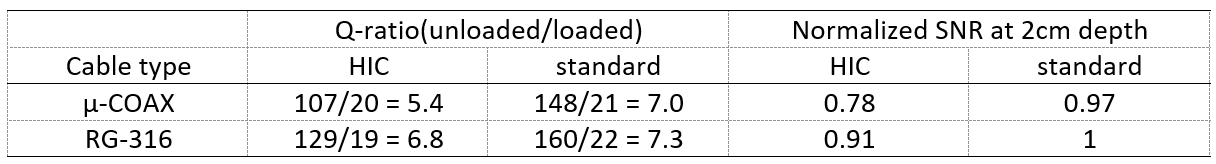
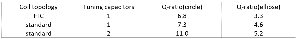
To explore flexible coil design options, we measured coil Q and SNR in 4 single-channel 7.5cm diameter test loops built from micro-coax or RG-316 and configured in “high impedance coil” (HIC) 5 or standard topologies 10. Next, we measured SNR in HIC and standard two-channel test arrays that were arranged with critical geometric overlap or with the conductor edges touching to replicate worst-case coupling. Finally, we measured Q when the coils were mechanically neutral (circular) or distorted into an ellipse, as would occur during knee flexion, with different numbers of distributed capacitors.Based on results from the experiments above, a flexible 6-channel array was constructed to wrap around the knee (Figure 1). The coils had conventional topology and were built using RG-316 coaxial cable and two tuning capacitors. While the natural coil dimensions (length = 14 cm and width = 9 cm) were calculated to accommodate the 50th percentile male knee size, the coil could be stretched laterally to accommodate the 90th percentile knee as well. This was made possible by the stretchable elastic shell (Fig. 1a) with integrated pockets designed to maintain approximately 20% overlap in the lateral direction between neighbor (Fig.1b) coils to maintain inductive decoupling.
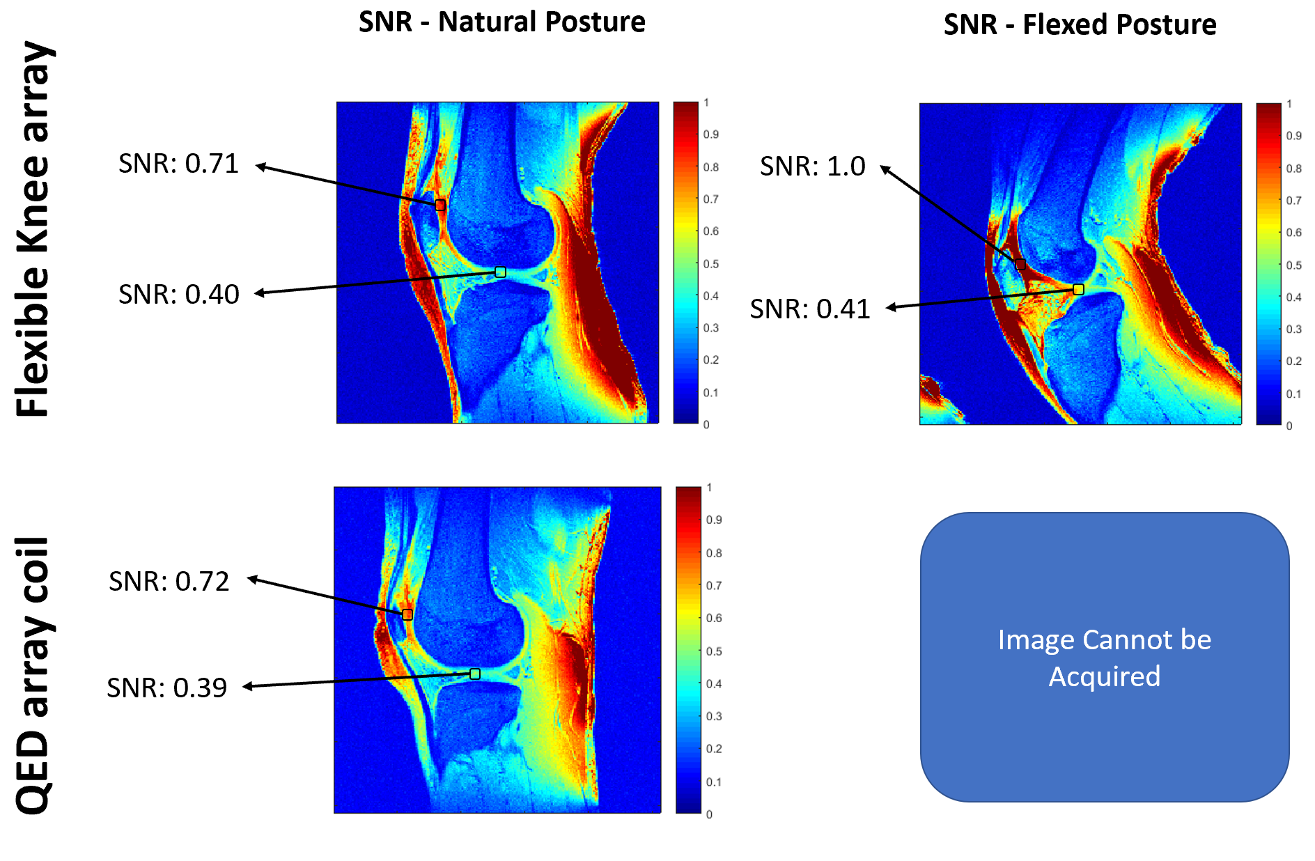
One subject was scanned on a 3T MRI system (Prisma, Siemens Healthcare, Erlangen, Germany) after providing informed written consent in accordance with our local internal review board. To demonstrate proof-of-concept, we measured SNR in the cartilage when the knee was at rest and flexed, and compared the measurements to those using a rigid commercial coil (15-channel knee array, QED, Mayfield, OH).
Results and Discussion
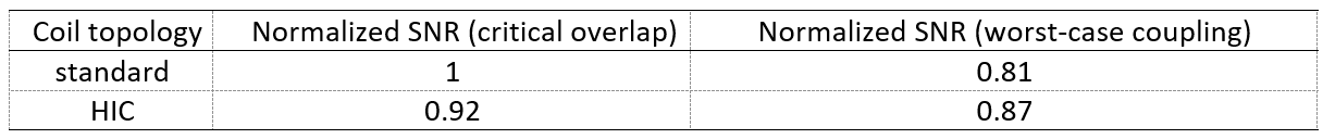
The single-channel Q and SNR tests revealed that the coil built from RG-316 with standard topology outperformed those with micro-coax or HIC topology (Table 1). Subsequent measurements focused only on coils built from RG-316, given that it provided adequate mechanical flexibility for our application and superior durability and lower loss compared to micro-coax.Table 2 shows that the standard coil topology with 2 distributed capacitors provided superior Q compared to that with 1 distributed capacitor or the HIC, with and without mechanical flexion. Table 3 shows that the two-channel array with standard topology outperformed the two-channel HIC array when the coils were critically overlapped, but underperformed when the coils were arranged for worst-case coupling.
Given these results, we built a 6-channel array from RG-316 with a conventional topology. The array was integrated into a flexible shell designed to allow knee flexion while preserving geometric decoupling that would otherwise compromise SNR: S21 of neighboring coils was -25.6 dB with the knee in a relaxed posture (Fig. 1c) and -17.5 dB during 40° flexion (Fig. 1d).
In vivo data show that respective SNR in the patellar (peripheral) and articular (central) cartilage in the relaxed posture were nearly identical with the proposed and commercial coils (Fig. 2). During 40° knee flexion, despite mechanical coil deformation, SNR with the proposed flexible array was maintained in the articular cartilage, and SNR improved by nearly 40% in the patellar cartilage compared to values measured in the natural posture. The 40° knee flexion posture was not possible with the rigid, commercial coil.
In conclusion, we built a flexible knee coil that enables MRI during knee flexion while providing similar or improved SNR compared to a state-of-the-art rigid commercial coil. The proposed coil was built from off-the-shelf coaxial cable and can be easily assembled with common tools. We anticipate that the coil will be useful for weight-bearing or kinetic knee imaging in which rigid coils provide suboptimal SNR and/or severely restrict the desired posture. While not demonstrated here, the wrap-around flexible array can accommodate various knee sizes, which may provide inherent SNR advantage over rigid coils 11.
Acknowledgements
The authors thank Jan Paska for insightful discussions. This work was partially supported by National Institutes of Health grants R21CA213169, R01DK106292, R21AG061579, R01DK114428, and R21EB027263 and was performed under the rubric of the Center for Advanced Imaging Innovation and Research (CAI2R, www.cai2r.net) at the New York University School of Medicine, which is an NIBIB Biomedical Technology Resource Center (NIH P41 EB017183).References
1. Bruno, F., et al., Weight-bearing MRI of the knee: a review of advantages and limits. 2018. 89(Suppl 1): p. 78.
2. Juras, V., et al., Kinematic biomechanical assessment of human articular cartilage transplants in the knee using 3-T MRI: an in vivo reproducibility study. 2009. 19(5): p. 1246-1252.
3. Mazzoli, V., et al., Accelerated 4D self‐gated MRI of tibiofemoral kinematics. 2017. 30(11): p. e3791.
4. Menon, R.G., M.V. Zibetti, and R.R. Regatte, In vivo tibiofemoral cartilage strain mapping under static mechanical loading using continuous GRASP‐MRI. 2020. 51(2): p. 426-434.
5. Zhang, B., D.K. Sodickson, and M.A. Cloos, A high-impedance detector-array glove for magnetic resonance imaging of the hand. 2018. 2(8): p. 570-577.
6. Ruytenberg, T., A. Webb, and I. Zivkovic, Shielded‐coaxial‐cable coils as receive and transceive array elements for 7T human MRI. 2020. 83(3): p. 1135-1146.
7. Port, A., et al., Detector clothes for MRi: A wearable array receiver based on liquid metal in elastic tubes. 2020. 10(1): p. 1-10.
8. Nordmeyer‐Massner, J.A., N. De Zanche, and K.P. Pruessmann, Stretchable coil arrays: application to knee imaging under varying flexion angles. 2012. 67(3): p. 872-879.
9. Nohava, L., et al. Flexible multi‐turn multi‐gap coaxial RF coils (MTMG‐CCs): design concept and bench validation. in Proceedings of the 27th Annual Meeting of ISMRM, Montreal, Canada. 2019.
10. Roemer, P.B., et al., The NMR phased array. 1990. 16(2): p. 192-225.
11. Zhang, B., et al., Size‐adaptable “Trellis” structure for tailored MRI coil arrays. 2019. 81(5): p. 3406-3415.
Figures