0129
Enhanced Ultra-High Field Brain MRI Using a Wireless Radiofrequency Sheet1Radiology, Icahn School of Medicine at Mount Sinai, New York, NY, United States, 2Neurosurgery, Icahn School of Medicine at Mount Sinai, New York, NY, United States, 3Radiology, University of Minnesota-Medical School, Minneapolis, MN, United States
Synopsis
Ultra-high field (UHF) MRI, such as 7T can visualize the brain in significantly improved detail through enhanced signal-to-noise ratio and contrast mechanisms. However, when the wavelength becomes comparable with the body dimensions, excitation radiofrequency (RF) field homogeneity at UHF systems is impaired by wavelength effects. Here we report a novel RF resonator sheet design with a simple circuit structure that couples inductively to the RF coil to enhance RF transmit homogeneity, efficiency, and signal sensitivity. In-vivo human experiment results demonstrate the feasibility and effectiveness of this method in brain MRI at 7T.
Introduction
The increased signal-to-noise ratio (SNR) and enhanced contrast available at ultra-high field (UHF) enable higher resolution anatomic, diffusion, functional, and vascular imaging1. However, one of the remaining problems is the excitation field ($$$B_1^+$$$) homogenization of the volume coil, which becomes challenging with the shorter RF wavelengths occurring at higher magnetic fields. As the RF wavelength becomes comparable with the body dimensions, the coil field becomes distorted. As a result, locations of destructive and constructive field interferences occur in sample, which results in dark voids and bright areas in MR images. Various active and passive RF-shimming techniques such as parallel transmission (PTx)2, coupling surface coils3, dielectric pads4, metasurfaces5, and artificial dielectric slabs6 were reported to tailor the at UHF. However, these methods have some drawbacks such as unstable material parameters of dielectric pads, high-cost and complexity of PTx systems, and complex structure of the metasurfaces. To achieve better transmit efficiency while avoiding the hardware complexity and other disadvantages of these prior methods, we propose a reliable passive RF resonator array that is based on an inductive-coupling between the passive RF sheet and the volume coil. We demonstrate the utility of the RF sheet in acquiring in-vivo human brain images focusing on the posterior fossa with enhanced local efficiency and signal sensitivity on a 7T MRI.Method
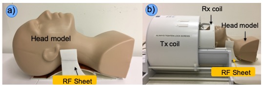
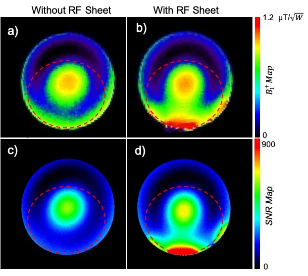
The fundamental principle behind this method is that transmit efficiency during the RF excitation and the receive signal sensitivity during the signal reception can be enhanced by the addition of an inductively coupled passive device placed near the anatomy of interest. The passive RF resonator sheet consists of an array of overlap-decoupled resonator elements, each 4.3 cm in diameter, patterned on a flexible substrate7. Each element is a multilayer structure consisting of two metal layers with a dielectric layer sandwiched in between. The structure size is 9 x 20 cm2 with a thickness of 200 μm and a 0.3 cm thick plastic foam on which covers copper layers. The flexible and thin structure of the RF sheet allows it to conform to curved surfaces to more effectively cover the target anatomy (Fig. 1a, b).We experimentally measured $$$B_1^+$$$ maps inside a head-mimicking phantom in the presence of the RF sheet and without the sheet. $$$B_1^+$$$ maps were measured using the double-angle method with the same input power a normalized by square roots of transmit RF power. To evaluate the enhancement of receive sensitivity, SNR maps were calculated by comparing two gradient-recall-echo (GRE) acquisitions, one with and the other without RF excitation.
An experimental temperature test was performed using an ASTM phantom for safety before human subject scans. Four fiberoptic temperature sensors were placed at locations with high energy deposition predicted by the simulation.
In-vivo MR images were obtained with/without the RF sheet in a human volunteer using GRE sequences (TR/TE=400/9 ms, FA=5°, bandwidth=977 Hz/pixel, FOV=9×14 cm2, matrix=256×256) with the transmit reference voltage kept constant. The RF sheet was placed in the inferior position behind the neck, which is associated with reduced transmit efficiency.
$$$B_1^+$$$ and SNR maps were calculated using the methods explained above. All experiments were performed on a 7T MRI scanner (Magnetom, Siemens Healthcare, Erlangen, Germany) using Nova Medical 1Tx/32Rx head coil.
Results
The spatial distribution of the $$$B_1^+$$$ maps in the experiments showed an improved performance toward the region-of-interest (ROI) when the RF sheet is present (Fig. 2a, b). In phantom experiments, the averaged across the ROI (outlined in dashed red) showed a mean 2-fold improvement in the presence of the RF sheet. Experimental phantom SNR maps showed an overall SNR enhancement of 2.4-fold in the presence of the RF sheet in the ROI (Fig. 2c, d).Base on the heating safety test, for safe scanning, the SAR monitor must not exceed 80% when the RF sheet is used.
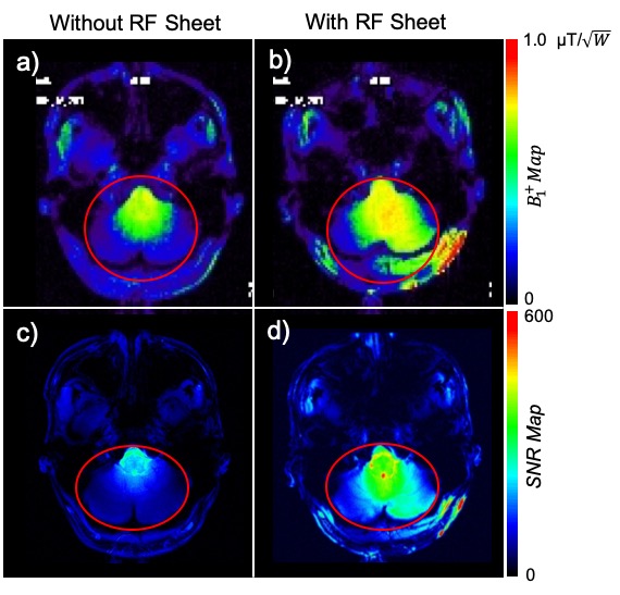
In-vivo $$$B_1^+$$$ and SNR maps showed a 1.7 and 2.0 enhancement-factors, respectively in the interested region (outlined in red) using the RF sheet (Fig. 3).
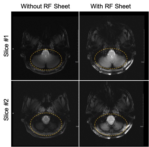
In-vivo brain MRI at 7T was conducted with the RF sheet positioned behind the neck, covering the posterior fossa. The T1-weighted axial low flip angle GRE images showed improved visibility of the cerebellum and the brainstem in the presence of the RF sheet (Fig. 4).
Discussion and Conclusion
This study has demonstrated a new design of a flexible and compact RF resonator sheet structure which can improve ultra-high field brain MRI in regions of lower transmit efficiency. In this demonstration we targeted the cerebellum, which is commonly studied in anatomical and functional experiments. Experimental results have shown that the RF sheet can enhance the local transmit field and improve the receive signal sensitivity at the periphery of an RF coil’s region of peak efficiency and sensitivity, which resulted in an average SNR enhancement of 2-fold in the posterior fossa. The proposed RF resonator sheet constructed of a very simple substrate can be considered a low-cost and time-stable alternative to other passive RF-shimming techniques at UHF MRI. We have demonstrated this method at 7T, but this technique can also be used at 3T and 1.5T. Future studies will focus on the optimization of the RF sheet under various standard MRI protocols.Acknowledgements
All studies involving human subjects were performed in accordance with the MRI safety review board in Icahn School of Medicine at Mount Sinai.References
[1] P. Balchandani and T. P. Naidich, "Ultra-High-Field MR Neuroimaging," vol. 36, no. 7, pp. 1204-1215, 2015.
[2] W.A. Grissom, L. Sacolick, M.W. Vogel, Improving high-field MRI using parallel excitation, Imag. Med. 2 (6) (2010) 675–693.
[3] Wang S, Murphy-Boesch J, Merkle H, Koretsky AP, Duyn JH. B1 homogenization in MRI by multilayer coupled coils. IEEE Trans Med Imaging. 2009 Apr;28(4):551-4. doi: 10.1109/TMI.2008.2006523. PMID: 19336276; PMCID: PMC3197805.
[4] Vaidya MV, Lazar M, Deniz CM, Haemer GG, Chen G, Bruno M, Sodickson DK, Lattanzi R, Collins CM. Improved detection of fMRI activation in the cerebellum at 7T with dielectric pads extending the imaging region of a commercial head coil. J Magn Reson Imaging. 2018 Aug;48(2):431-440. doi: 10.1002/jmri.25936. Epub 2018 Jan 21. PMID: 29357200; PMCID: PMC6054823.
[5] Schmidt, R., Slobozhanyuk, A., Belov, P. et al. Flexible and compact hybrid metasurfaces for enhanced ultra high field in vivo magnetic resonance imaging. Sci Rep 7, 1678 (2017). https://doi.org/10.1038/s41598-017-01932-9
[6] Vorobyev V, Shchelokova A, Zivkovic I, Slobozhanyuk A, Baena JD, Del Risco JP, Abdeddaim R, Webb A, Glybovski S. An artificial dielectric slab for ultra high-field MRI: Proof of concept. J Magn Reson. 2020 Nov;320:106835. doi: 10.1016/j.jmr.2020.106835. Epub 2020 Sep 24. PMID: 33065392.
[7] Alipour, A., Seifert, AC., Delman, D., Adriany, G., Balchandani P. Improvemnet of brain MRI at 7T using an inductively coupled RF resonator array. 2020, arXiv preprint arXiv:2006.02500.
Figures