0122
Motion Estimation for Brain Imaging at Ultra-High Field Using Pilot-Tone: Comparison with DISORDER Motion Compensation1Biomedical Engineering Department, School of Biomedical Engineering and Imaging Sciences, Kings College London, London, United Kingdom, 2Centre for the Developing Brain, School of Biomedical Engineering and Imaging Sciences, Kings College London, London, United Kingdom, 3MR Research Collaborations, Siemens Healthcare Limited, Frimley, United Kingdom, 4Biomedical Image Technologies, ETSI Telecomunicación, Universidad Politécnica de Madrid and CIBER-BNN, Madrid, Spain
Synopsis
A ‘pilot-tone’ implementation for 7T head MRI was constructed by broadcasting RF at the appropriate frequency into the scanner room during data acquisition. This signal was demonstrated to enable motion estimation, when calibrated first by correlating measurements with motion estimates from image registration. Subsequently these estimates were compared with others obtained from the iterative DISORDER joint motion estimation & reconstruction method. These independent methods of motion estimation can potentially improve or replace other methods of motion correction at ultra-high field where motion-correction is particularly relevant.
Introduction
Motion artefacts in the head are well documented1 and have a significant impact on high resolution image quality, particularly relevant for ultrahigh field imaging.2DISORDER is a retrospective motion-correction method that compensates for rigid body motion during 3D acquisitions,3,4 essentially by simultaneously estimating intra-scan motion parameters jointly with the reconstructed images.
Pilot-tone is a motion-estimation method that uses an externally generated RF signal consisting of a single frequency chosen such that it falls outside the field of view of an image but is still sampled in the k-space data, within the over-sampled region of the frequency encoded direction.5 It has been demonstrated6 for abdominal imaging at lower field strengths, that respiration-induced loading changes of a local array receiver coil will cause temporal fluctuations in this pilot-tone signal that can then be used to infer motion information. In this work we have explored use of a similar method to create head motion estimates for brain imaging at 7T, and compare these estimates with those from the DISORDER framework.
Methods
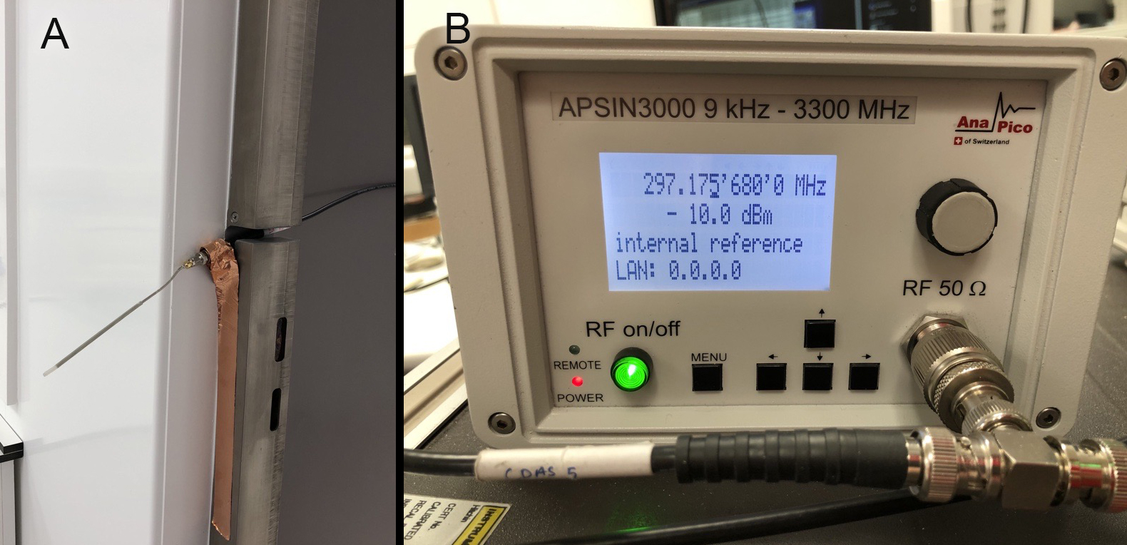
All measurements were performed on a MAGNETOM Terra (Siemens Healthcare, Erlangen, Germany) 7T scanner in research configuration, with an 8-Tx-channel head coil (Nova Medical, Wilmington MA, USA). Human subject scanning was approved by the Institutional Research Ethics Committee (HR-18/19-8700). All scans were carried out on a single, healthy volunteer. All data analysis was carried out using MATLAB R2020b (MathWorks, Natick, MA, USA).A pilot-tone was generated with an RF signal generator (APSIN3000, AnaPico, Glattbrugg, Switzerland) with an offset frequency in the over-sampled frequency-encoded data (adjusted per scan). The tone was piped into the scanner room through a waveguide via a monopole antenna (Figure 1), the power level set on the signal generator was –10.0dBm.
An initial set of 40 GRE “calibration” scans were acquired with the volunteer instructed to hold a different static position during each measurement, to provide positional references for the pilot tone data. GRE hold protocol: TE/TR = 5/10 ms; flip angle: 7°; 220x110x112 matrix (ROxPE1xPE2), 240x240x112 mm3 field of view (FOV); GRAPPA 2x2.
These were then followed by a DISORDER GRE scan with the volunteer instructed to provide a variety of different movements throughout.3 DISORDER protocol: TE/TR = 5/10 ms; flip angle: 7°; 220x110x112 matrix (ROxPE1xPE2), 230x230x112 mm3 FOV.4
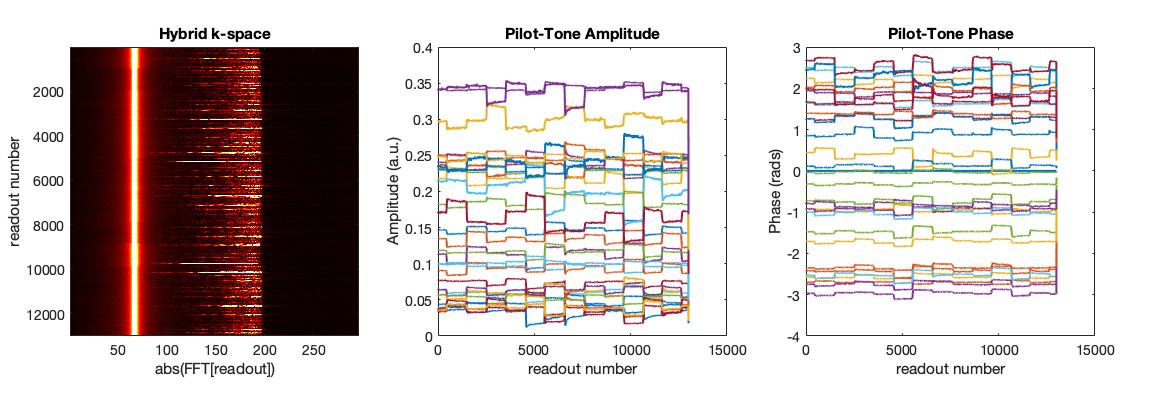
A hybrid k-space5, containing the pilot-tone, was generated by applying a Fourier transformation to each readout line in the acquired k-space. The amplitude of the detected peak in the oversampled region was selected as the pilot-tone. Then the pilot-tone phase was referenced to the first coil and the amplitude was normalized across coils. Finally, a median filter with a 10-sample window was applied.
Motion parameters from the DISORDER reconstruction were obtained using the joint optimisation previously proposed.3
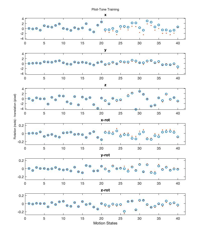
Motion parameters for the calibration data were estimated using rigid-body registration of the 40 GRE images (39 images registered to the first volume). The pilot-tone signals $$$x$$$ were assumed to relate to the registration parameters $$$b$$$ via linear model $$$Ax=b$$$. This model could be calibrated by estimating $$$A$$$ from measurements where $$$b$$$ is known (via registration) computing $$$A=bx^{-1}$$$ using the pseudo-inverse to invert the under-determined $$$x$$$, aiding the estimate of motion parameters for any new pilot-tone measurement. We experimented with using either magnitude or complex data for $$$x$$$ and found that magnitude only data was better able to generalise to unseen measurements.
After fitting the linear model, the predicted motion parameters from the pilot-tone signal were provided to the DISORDER reconstruction. First, images were reconstructed using only pilot-tone motion estimates (no iterative motion estimation). Lastly, pilot-tone motion traces were used to guide data driven estimation of pose dependent B0-fields using a recently proposed extension of the DISORDER reconstruction8.
Results
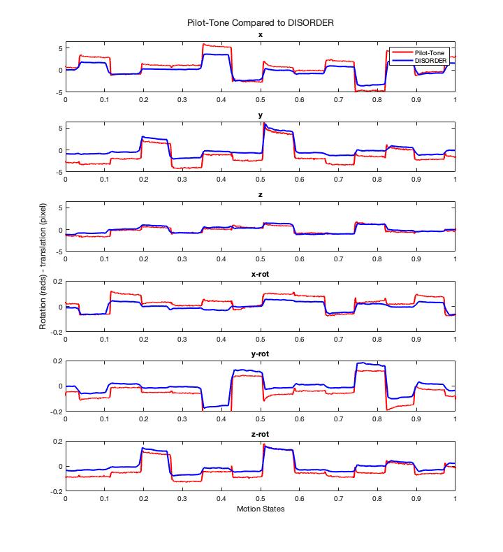
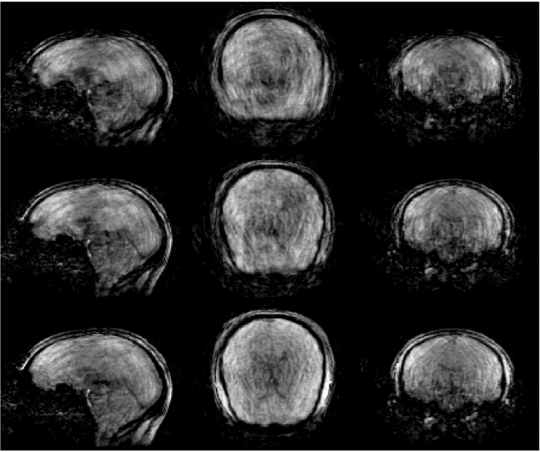
Figure 2 shows the pilot-tone in hybrid k-space and plots of the extracted amplitude and phase for each coil. Figure 3 shows the calibration of the motion estimation model from pilot tone data trained from the first 20 GRE images and then tested on the second set of 20 GRE images. Figure 4 shows pilot-tone motion estimates compared with DISORDER motion estimates. Figure 5 shows DISORDER reconstructed images. The top row shows uncorrected data, the middle row data corrected with the pilot-tone motion estimates, the bottom row corrected data using the pilot-tone motion estimates to compute pose-dependent fields.Discussion
This experiment describes first experience of using a pilot-tone to estimate head motion, at ultra-high field. Results indicate that the use of a monopole antenna with low-power pilot-tone signal at the entrance to the room is enough to generate useful motion estimates from a 32-channel receiver. Once trained, the motion model was able to accurately predict translation and rotation parameters only from the pilot-tone data.The motion estimates were compared with others that come directly from the DISORDER joint motion estimation/reconstruction method and are in good qualitative agreement, though some differences exist.
Conclusion
We have shown that head-motion estimates can be obtained from pilot-tone measurements at ultra-high field and correlated them to motion estimation from the DISORDER method. Independent motion estimates of this type could potentially be used to augment or replace other motion correction methods. Future work would focus on an efficient method for in situ calibration.Acknowledgements
The authors would like to thank Francesco Padormo for the loan of the RF signal generator.
This work was supported by core funding from the Wellcome/EPSRC Centre for Medical Engineering [WT203148/Z/16/Z] and by the National Institute for Health Research (NIHR) Biomedical Research Centre based at Guy’s and St Thomas’ NHS Foundation Trust and King’s College London and/or the NIHR Clinical Research Facility. The views expressed are those of the author(s) and not necessarily those of the NHS, the NIHR or the Department of Health and Social Care.
References
1. Andre JB, Bresnahan BW, Mossa-Basha M, et al. Toward Quantifying the Prevalence, Severity, and Cost Associated With Patient Motion During Clinical MR Examinations. J Am Coll Radiol. 2015;12(7):689-695. doi:10.1016/j.jacr.2015.03.007
2. Zaitsev M, Maclaren J, Herbst M. Motion artifacts in MRI: A complex problem with many partial solutions. Journal of Magnetic Resonance Imaging. 2015;42(4):887-901. doi:10.1002/jmri.24850
3. Cordero-Grande L, Ferrazzi G, Teixeira RPAG, O’Muircheartaigh J, Price AN, Hajnal JV. Motion-corrected MRI with DISORDER: Distributed and incoherent sample orders for reconstruction deblurring using encoding redundancy. Magnetic Resonance in Medicine. 2020;(November 2019):1-14. doi:10.1002/mrm.28157
4. Cordero-Grande L, Tomi-Tricot R, Ferrazzi F, Sedlacik J, Malik SJ, Hajnal JV. Preserved high resolution brain MRI by data-driven DISORDER motion correction. In: Proc Int Soc Mag Reson Med 20.
5. Bacher M. Cardiac Triggering Based on Locally Generated Pilot-Tones in a Commercial MRI Scanner: A Feasibility Study. October 2017.
6. Vahle T, Bacher M, Rigie D, et al. Respiratory Motion Detection and Correction for MR Using the Pilot Tone. Investigative Radiology. 2020;55(3):7.
7. Cordero-Grande L, Teixeira RPAG, Hughes EJ, Hutter J, Price AN, Hajnal JV. Sensitivity Encoding for Aligned Multishot Magnetic Resonance Reconstruction. IEEE Transactions on Computational Imaging. 2016;2(3):266-280. doi:10.1109/tci.2016.2557069
8. Brackenier Y, Cordero-Grande L, Tomi-Tricot R, et al. Data-driven motion-corrected brain MRI incorporating pose dependent B0 fields. In: Proc Int Soc Mag Reson Med 21. (Submitted to ISMRM 2021)
Figures