0078
Fluorine Magnetic Resonance Imaging for Natural Killer Cell Tracking with a Dual Tuned 1H/19F Torso Coil at 3T1Medical Physics, Carbone Cancer Center, University of Wisconsin School of Medicine and Public Health, Madison, WI, United States, 2Pediatrics, Carbone Cancer Center, University of Wisconsin School of Medicine and Public Health, Madison, WI, United States, 3Medical Sciences, School of Veterinary Medicine, University of Wisconsin-Madison, Madison, WI, United States, 4Carbone Cancer Center, University of Wisconsin-Madison, Madison, WI, United States, 5Radiology, Carbone Cancer Center, University of Wisconsin School of Medicine and Public Health, Madison, WI, United States, 6Biomedical Engineering, Carbone Cancer Center, University of Wisconsin School of Medicine and Public Health, Madison, WI, United States
Synopsis
Fluorine magnetic resonance imaging (19F-MRI) has been demonstrated as a non-invasive method to track and quantify immune cells in vivo. However due to the low 19F spin density of immune cell labeling, these studies have been mostly conducted on ultra-high field MRI systems, or with small sensitive surface coils at clinical field strengths. This feasibility study found that concentrations of perfluoropolyether (PFPE), and phantoms consisting of fewer than one million PFPE labeled NK cells were reliably detected through 19F-MRI with the combination of a cartesian 3D fast spin echo imaging sequence, and a dual tuned 1H/19F torso coil at 3T.
Introduction
Fluorine magnetic resonance imaging (19F-MRI) has been demonstrated as a non-invasive method to label, track and quantify T-cells, natural killer cells (NK cells) or genetically altered chimeric antigen receptor T-cells (CAR T) in vivo, which are evolving as novel immunotherapy cancer treatments [1-3]. However due to the low spin density of 19F nuclei obtained through immune cell labeling in vivo; to date, these studies have been mostly conducted on ultra-high field (B0≥7T) small animal MRI systems, or with small sensitive surface coils at clinical field strengths, which are not ideal for whole body in vivo cellular tracking [1-3]. Therefore, this pilot study set out to investigate the feasibility of NK cell tracking on a clinical 3T MRI with a dual tuned 1H/19F torso coil, through the investigation of 19F labeled NK cell phantoms measurements, which will lay the groundwork for future in vivo studies in canine osteosarcoma patients.Methods
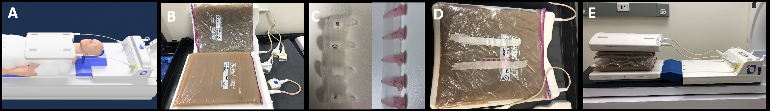
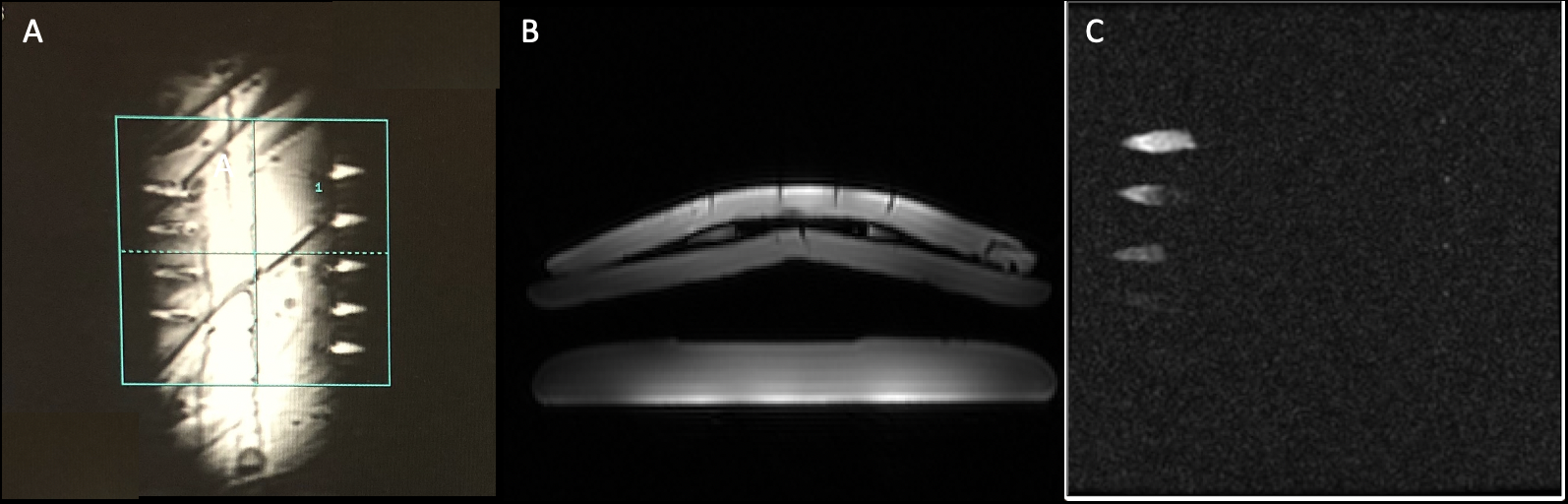
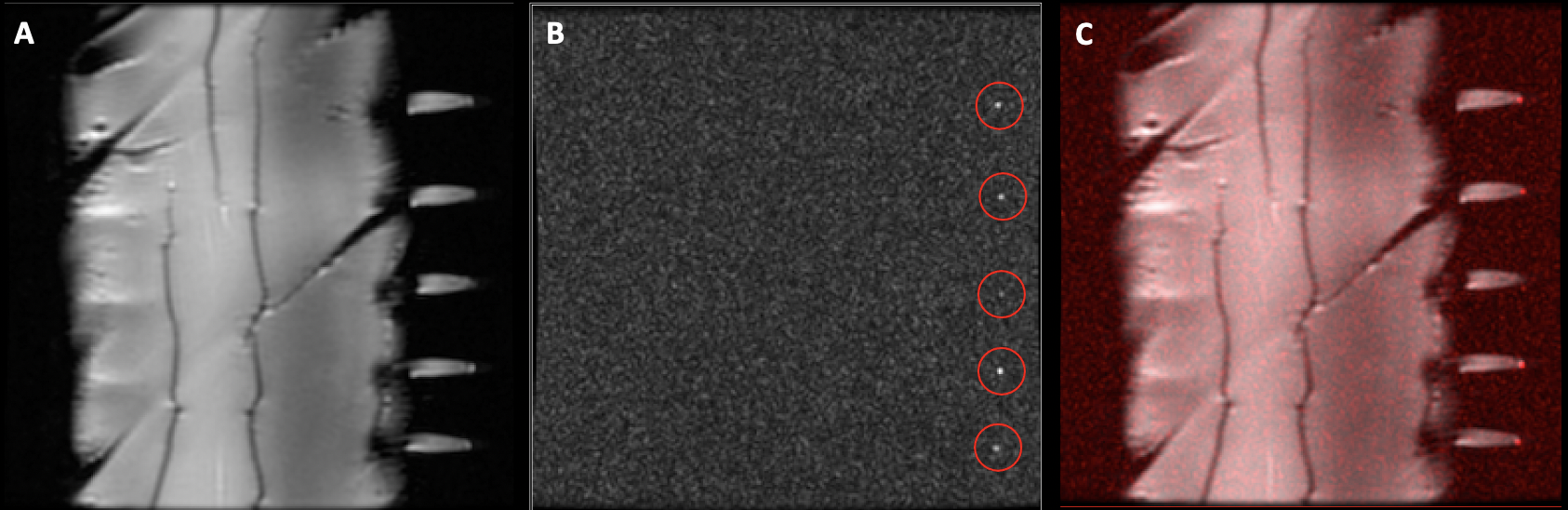
All measurements were conducted on a 3.0T Discovery MR750w MRI scanner (GE Healthcare, Waukesha, WI) with a 8-channel dual tuned 1H/19F torso coil (MRI Tools, Berlin, Germany) (Figure 1A). Reproducibility phantom measurements (N=3) were acquired on separate days, with a cartesian 3D fast spin echo sequence (TR: 750ms, TE: 27.4ms, ETL: 16, FOV: (200 x 200 x 42)mm, Matrix: 128 x 128, resolution: (3 x 1.6 x 1.6)mm, BW: 14.71kHz, NEX: 32, T = 45 min). Agar phantoms (2% by weight, KCL: 30mM) were made which covered the surfaces of the coil elements, to ensure proper loading and spacing during phantom measurements (Figure 1B), and a 0.5 ml sample of pure Perfluoro-15-crown-5 ether (Exfluor, Round Rock, Texas) was used for manual flip angle calculation. Phantoms used for the feasibility study consisted of perfluoropolyether (PFPE) phantoms with 25ul, 50ul, 100ul, 200ul of CS-1000 (Celsense Inc, Pittsburgh, PA) homogenously distributed in 1.5 ml agar vials, and canine NK cell phantoms (#cells: 0.3x10⁶, 0.6x10⁶, 1.2x10⁶, 3x10⁶, 6x10⁶) which were co-cultured with 8.0 mg/ml CS-1000 for 4-Hrs (Figure 2C). The canine NK cells were pelleted at the bottom of a 2 ml conical tube by centrifugation and filled with agar as described previously [3]. Cell viability of 60%, and a labeling efficiency of 3.65x10¹² (19Fnuclei/cell) were assumed from previous results in our lab [2]. Additional agar loading phantoms were used to position the PFPE phantoms and simulate the loading and spacing of a typical canine body (Figure 2 D-E). Signal-to-noise of all images was measured in FIJI [4], as calculated as the mean of the signal over the standard deviation of the noise.Results
Cartesian 3D fast spin echo 19F-MRI of the homogenous PFPE phantoms across (N=3) days revealed a increase in SNR corresponding with total volume of PFPE [25ul: 7.1±0.6 (mean±SD), 50ul: 13.4±2.0, 100ul: 21.8±1.9, 200ul: 31.7±5.2) within the 45-min scan (Figure 2). In addition to this, the pelleted PFPE labeled NK cell phantoms were reproducibly detected with a low variation in SNR at a resolution of (1.6x1.6x3.0)mm during each imaging session (Figure 3). However, either due to phantom construction or NMR sensitivity, the small point NK cell phantoms were detected with a comparable SNR across all quantities of PFPE labeled NK cells [0.3x10⁶: 13.3±2.0, 0.6x10⁶: 13.9±3.0, 1.2x10⁶:11.9±4.5, 3x10⁶:16.4±3.4, 6x10⁶:14.1±3.8).Conclusion
This feasibility phantom study found that concentrations of PFPE were reliably detected with increased SNR through 19F-MRI with the combination of a cartesian 3D fast spin echo imaging sequence, and a dual tuned 1H/19F torso coil at 3T. More importantly, phantoms consisting of fewer than one million PFPE labeled NK cells were also consistently detected with the dual tuned torso coil at a similar distance and RF-loading to those expected in an in vivo canine osteosarcoma patient. Nevertheless, these results show that additional improvements in pulse sequence development, and in vivo cell quantification need to be investigated, including the investigation of non-cartesian 3D k-space sampling techniques [5] which should lead to improved scan efficiency and sensitivity to PFPE labeled NK cells in vivo.Acknowledgements
-
This project used shared facilities
provided in part by the University of Wisconsin, Department of Medical Physics
and the UW-Carbone Comprehensive Cancer Center
- This project was supported by a radiological sciences training grant T32 CA009206 fellowship to Dr. Paul Begovatz
- This project was supported in part by Celsense
- This project was supported by the ongoing collaboration of GE Healthcare with the University of Wisconsin-Madison
References
1. Srinivas M, Heerschap A, Ahrens E.T, et al., 19F MRI for quantitative in vivo cell tracking. Trends in Biotechnology 2010; 28:363-370.
2. Bouchlaka M, Ludwig K, Gordon J et al.19F-MRI for monitoring human NK cells in vivo. Oncoimmunology. 2016; 5:e1143996.
3. Ahrens E.T, Helfer B.M, O’Hanlon C et al. Clinical Cell Therapy Imaging Using a Perfluorocarbon Tracer and Fluorine-19 MRI. Magn. Reson. Med. 2014; 72:1696-1701.
4. Schindelin, J.; Arganda-Carreras, I. & Frise, E. et al. (2012), "Fiji: an open-source platform for biological-image analysis", Nature methods. 2019; 9(7): 676-682
5. Floegel U, Ahrens ET. Fluorine Magnetic Resonance Imaging. 2017 Pan Stanford Publishing Pte. Ltd.
Figures