S98
Fetal Cardiac Imaging Utilizing Retrospectively Gated 2D Radial MRI1Radiology, Boston Children's Hospital, Boston, MA, United States, 2Boston Children's Hospital, Boston, MA, United States, 3Advanced Clinical Imaging Technology, Siemens Healthcare, Lausanne, Switzerland, 4Radiology, Lausanne University Hospital and University of Lausanne, Lausanne, Switzerland, 5LTS5, Ecole Polytechnique Federale de Lausanne (EPFL), Lausanne, Switzerland, 6LTS5, École Polytechnique Fédérale de Lausanne (EPFL), Lausanne, Switzerland
Synopsis
Fetal MRI is a rapidly growing field that facilitates diagnosis and
treatment providing improved maternal and fetal care. New imaging strategies
are needed to improve the assessment of cardiac structure and function. We assessed
an interactive fetal cardiac prototype sequence enabling the fetal heart to be imaged in real-time, while prescribing the imaging plane to fetal movement. Once positioned to the long
axis, a sequence of 2D radial readouts are acquired, enabling inline
reconstruction of beating heart images with a frame rate between 0.5s
and 1s. This new capability allowed assessment the anatomy and function of
the fetal heart.
Background
Magnetic Resonance Imaging is an important tool in providing advanced fetal care. Since the first fetal MRI exam reported in 1985, this discipline has grown exponentially assisting obstetricians and neonatologists in confirming fetal and placental anatomy which enables early in-vitro and neonatal intervention [1]. Ultrasound has been the gold standard for fetal cardiac imaging, but it encounters issues due to fetal movement and maternal body habitus. For patients for whom ultrasound is unable to provide a clear answer, referral to MRI is common due to its improved ability to provide an accurate diagnosis [2]. Fetal cardiac imaging in MRI has been a focus of technological development, with many methods being trialed, ranging from fast sequences to new strategies for fetal cardiac gating. Similar to ultrasound, fetal cardiac imaging in MRI has its own obstacles from fetal and maternal motion, the length of exams, and limited spatial and temporal resolution [3]. Here, we assessed a fetal cardiac prototype sequence with interactive real-time capabilities (Siemens Healthcare, Erlangen, Germany) [5].Method
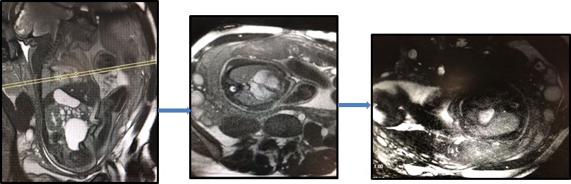
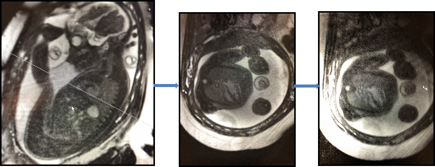
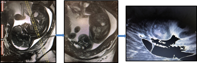
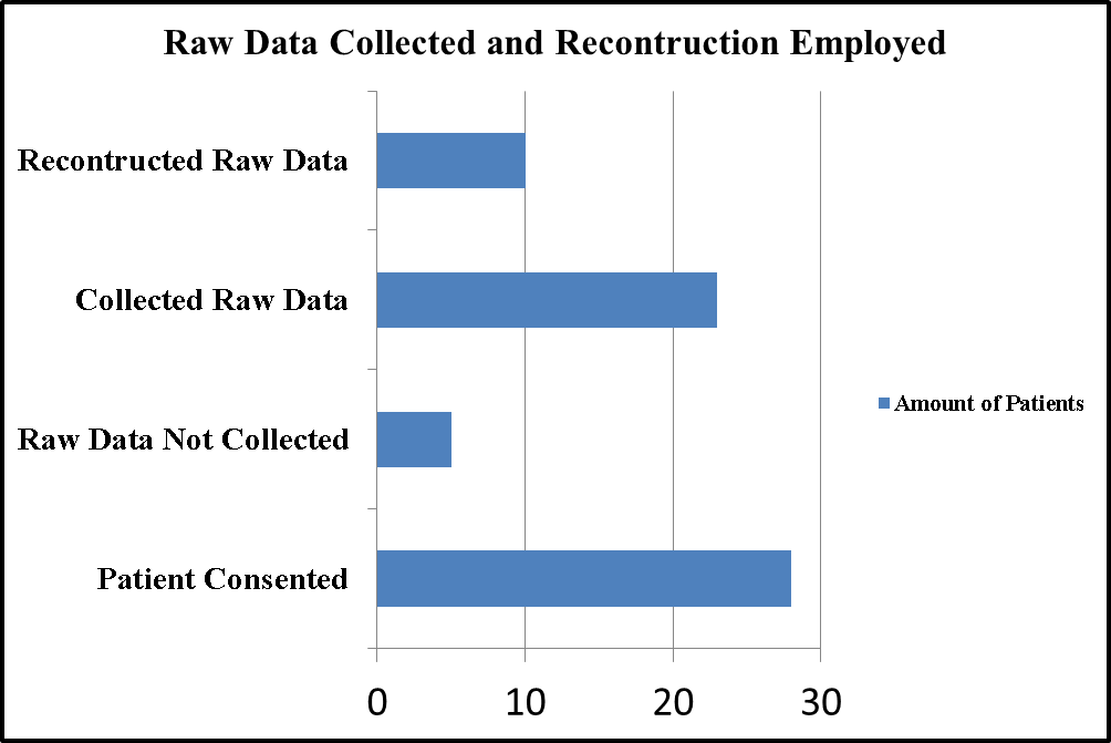
In 2019 our institution performed over 600 fetal MRI exams. With our ever increasing patient volume, 7.5% from 2018-2019, and our large cohort of infants born with congenital heart disease, there is a need for rapid and effective evaluation of the fetal heart. Therefore, we sought to evaluate an interactive fetal cardiac prototype sequence. The patient recruitment process consisted of identifying and offering participation to every subject meeting the enrollment criteria of a singleton pregnancy and being referred for a diagnostic fetal MRI exam. Written informed consent to participate in a research study approved by our IRB was obtained in every subject who agreed to volunteer. All exams were performed using a 30 channel body matrix at 3T (MAGNETOM Skyra, Siemens Healthcare, Erlangen, Germany) with the patient supine or lateral decubitus. The imaging volume was prescribed off a prior HASTE image. Once the heart was located (Figure 1, image 1), a single 2D radial slice was prescribed to acquire a long axis image. With the real-time capability of the prototype, we were able to rotate and move the imaging plane in real-time while imaging any plane of the heart. Once the plane that best visualizes the flow of the heart was determined, successful reconstruction of the MRI fetal heart requires no bulk motion for 10 seconds in order to collect enough data for a GRASP (Golden-angle RAdial Sparse Parallel) reconstruction. Not every fetus will hold still for any period of time can be difficult task. Once 10 seconds is achieved the sequence is stopped. The raw data is then manually reconstructed with an offline GRASP reconstruction [6]. Reconstruction of the raw data on the scanner required 5-10 minutes after each participant’s exam. Some of the reconstructed images displayed a streaking artifact which suggests an insufficient number of radial lines were acquired (Figure 3).Results
Each participant was consented under an appropriate IRB from our institution. Participants consisted of 28 female outpatients ranging between the gestational ages of 18 to 36 weeks averaging 22 weeks. Out of 28 consented patients we were able to successfully collect raw data files from 23 patients. The raw data was then loaded into the GRASP reconstruction application resulting in 10 reconstructed data sets displaying the heart beating. The IRT images were transferred to the patient’s medical record for radiologist and physician review when the GRASP reconstruction failed.Conclusion
With the growing need of fetal MRI for diagnosis and in-utero or neonatal interventions, there is a growing interest to provide more imaging techniques that are adapted to the motion of the fetus and mother. The fetal cardiac prototype sequence has allowed us to visualize the beating heart of the fetus, offering new information about the structure and function of the heart. A helpful feature contributing to the success of the prototype is the interactive real-time slice prescription mode. Allowing technologists to follow the movement of the fetus in order to capture the heart in real-time is an innovative tool which could be investigated in other imaging situations. As this prototype application technology matures, we expect the success rate will increase. This will enable the assessment of cardiac structure and function to become part of routine clinical care.Acknowledgements
No acknowledgement found.References
1. G. Gorincour, B. B.-N. (2007). Feasibility of Fetal Cardiac Magnetic Resonance Imaging: Preliminary Experience. Ultrasound Obstet Gynecol, 105-108.
2. L. Manganaro, V. V. (2014). Magnetic Resonance Imaging of Fetal Heart: Anatomical and Pathological Findings. The Journal of Maternal-Fetal & Neonatal Medicine, 1213-1219.
3. C. Garet, H. B. (1998). Magnetic Resonance Imaging of the Fetus. Pediatr Radiol, 201-211.
4. M. Fogel, R. D. (2005). Preliminary Investigations into a New Method of Functional Assessment of the Fetal Heart Using a Novel Application of "Real-Time" Cardiac Magnetic Resonance Imaging. Fetal Diagn Ther, 475-480.
5. Piccini D, Yerly J, Chaptinel J, et al. As Easy as Echo : Interactive Fetal Cardiac MR Imaging, ISMRM 2017
6. Chaptinel, J., Yerly, J., Mivelaz, Y. et al. Fetal cardiac cine magnetic resonance imaging in utero . Sci Rep 7, 15540 (2017) doi:10.1038/s41598-017-15701-1
Figures