S97
A technical approach to reduce inhomogeneities and image distortion in PET/MRI imaging using mMR Breast coil1Molecular Imaging & Nuclear Medicine, Indraprastha Apollo Hospitals, New Delhi, India, 2Physics, Vivekananda Global University, Jaipur, India, 3PET SUITE, House of Diagnostics, New Delhi, India
Synopsis
Inhomogeneity in fat suppression and image distortion pose problem in Breast MRI specially in the diffusion and dynamic fat suppressed images using mMR breast coil. The purpose of this work to improve fat-superstation and minimize image distortion in the breast imaging before applying the MRI sequence specially in Dynamic fat-supressed Contrast and Diffusion images. 330 breast cancer patients have been taken for this study. Volume shim was applied in all patients and water peak shift corrected manually when required. Our results show reduction in image distortion in diffusion and improving in fat-supressed MRI images.
Introduction
Dynamic contrast Enhanced breast MRI is a gold standard Imaging modality to differentiate between benign and malignant lesion. Inhomogeneity in fat suppression and image distortion pose problem in Breast MRI specially in the diffusion and dynamic fat suppressed images using mMR breast coil (1,2). Presence of non-uniformity in the radio-frequency (RF) coil pose major concerns. and several methods were used like segmentation and registration to reduce inhomogeneities and image distortion.Aim
The aim of the work is to examine effects of selective volume shim and correction of the water peak prior to image acquisition to improve fat-superstation and minimize image distortion in the breast imaging.Material and Methods
Three hundred thirty (330) breast cancer patients who underwent PET/MRI imaging form April 2018 to October 2019 using Simultaneous PET/MRI (Biograph mMR, Siemens) with a dedicated breast coil form the material of this study. Volume shim was applied in all patients and in case of water peak shift which caused improper fat-suppression and image distortion were corrected with manual water peak adjustment before acquiring MRI sequences.Results
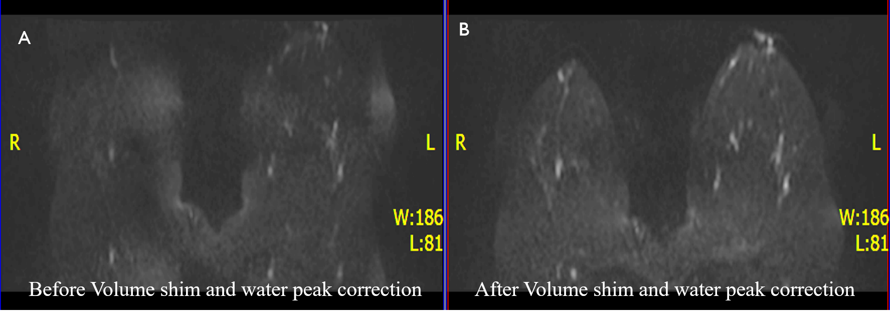
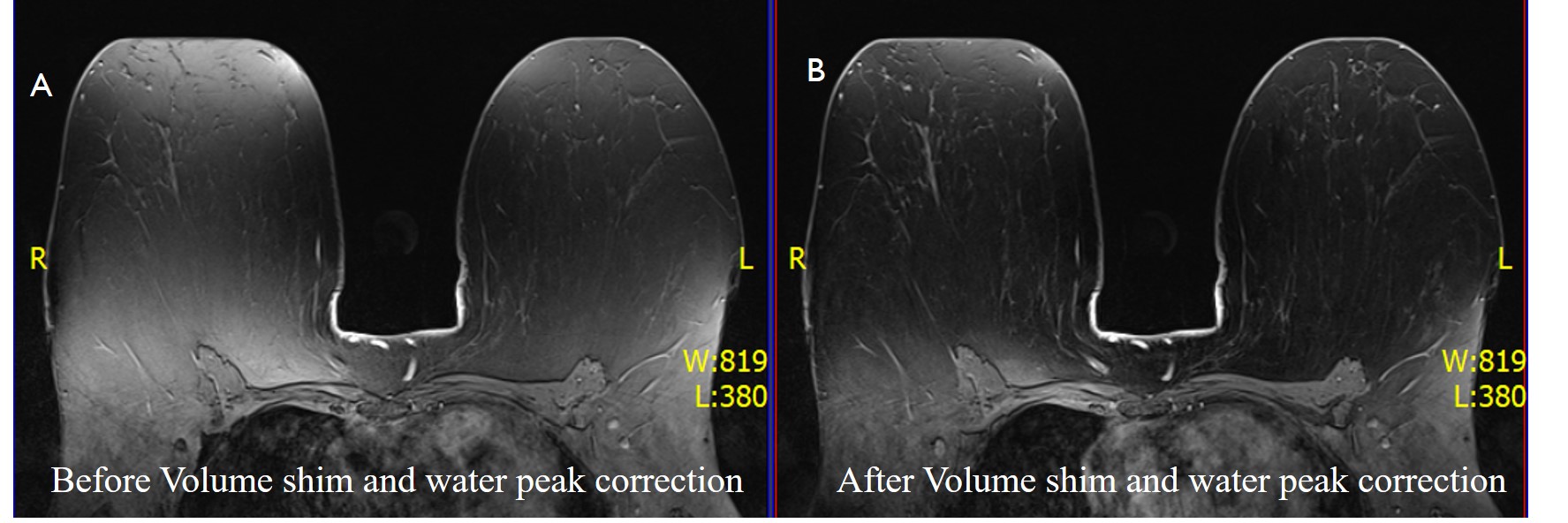
Our results show reduction in image distortion in diffusion imaging and improving homogeneity in dynamic fat-supressed contrast breast MRI ( Figure 1 ). Additional 15 sec was spent for water peak correction with no extra time being spent for volume shim. The protocol yields improving diagnostic image quality and lesion detection in the breasts MRI without repeating MRI sequence.Conclusion
Volume shim and water peak correction improves uniformity of fat suppression and reduce image distortion in breast MRI and may be included in the imaging protocol as routine.Key words
Breast MRI, Inhomogeneity, selective volume shim and water peak.Acknowledgements
No acknowledgement found.References
1. B1 Field correction of T1 estimation should Be considered for Breast Dynamic contrast-enhanced MR imaging even at 1.5 T1Wan-Chen Tsai, MD Kuo-Jang Kao, MD, PhD Kai-Ming Chang, PhD Chen-Fang Hung, MS et al, Radioloy 2017.
2. Federico D Pineda, Milica Medved, Xiaobing Fan et. Al. “B1 and T1 mapping of the breast with a reference tissue method” Magn Reson Med. 2016 April; 75(4): 1565–1573.
Figures