S86
Fat Fraction may be used as a radiographic evaluation index for vertebral bone metastasis:a report of 4 cases1Department of Radiology, The first affiliated hospital of xi 'an jiaotong university, Xi'an, China, 2MR Research China, GE Healthcare, BeiJing, China
Synopsis
Bone metastasis is a common complication of advanced malignant tumors with severe simptoms. Common imaging modalities for bone metastasis diagnosis include X-ray, CT,MRI, SPECT, and PET. PET is the golden standard, but the nature of radiation, metabolic risk and high price have limited PET from widely clinical use. In this work, we presented 4 cases of bone metastasis which had had a difference in MRI derived fat fraction between bone metastatic vertebrae and normal non-metastatic vertebrae. MR results were also coincided with PET results. MRI derived fat fraction may be used as an imaging evaluation index for bone metastasis.
Introduction
Common complications of skeletal tumor disease include bone metastasis, and the primary cancers that are most prone to spine metastasis are breast cancer, lung cancer, prostate cancer and kidney cancer. The location of 70% incidence of bone occurred in the thoracic vertebra, followed by the lumbar vertebra and cervical vertebra 1-2. At present, commonly used examination techniques for vertebral metastasis include CT, MRI, SPECT, and PET. CT and MRI are anatomical imaging techniques that can analyze tumor tissues on the basis of their morphological appearance while 18FDG PET and SPECT are functional imaging techniques. PET can differentiate malignant tumors, benign tumors and normal tissues, with diagnostic accuracy in the application of spinal metastatic tumors reported to be higher than 90%3-4. However, PET or SPECT requires tracer administration. For example, 18F-FDG required by PET is radioactive. Metabolic and radioactive contamination is a concern. Besides, to date, availability, accessibility, and affordability are still major limitations for PET examination. Recently presented MR technique, termed as IDEAL-IQ, can obtain tissues proton density fat fraction (PDFF) with high spatial resolution. IDEAL-IQ has been applied to various musculoskeletal examinations along with traditional magnetic resonance morphological imaging technology. The purpose of this study is to report several cases in the author's daily work that Fat Fraction of IDEAL -IQ technical might help with the diagnosis of bone metastasis.Methods
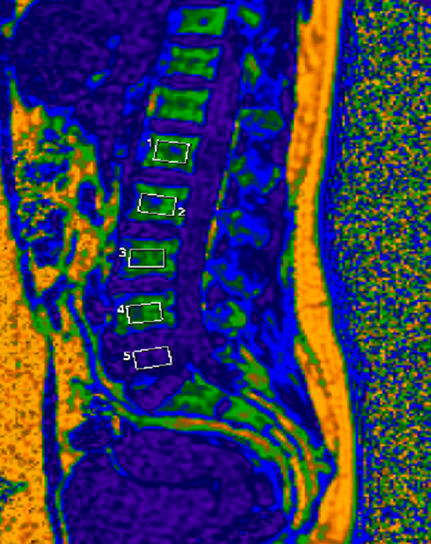
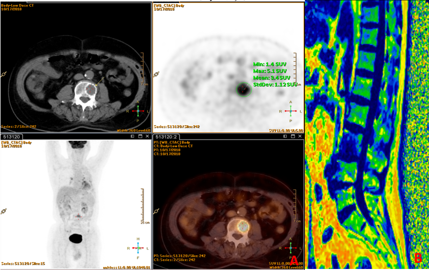
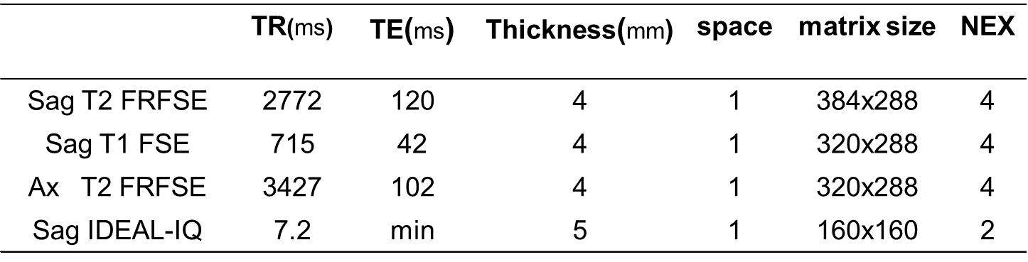
In this study, we collected a total of four patients with suspected vertebral bone metastasis. One case of prostate cancer, one case of lung cancer and two cases of breast cancer. All participants gave written informed consent approved by the local Research Ethics Committee after a complete description of this study. All subjects underwent PET, MR and SPECT scan. MR examinations were performed on a 3T MR scanner (Discovery MR750W , GE Healthcare, Milwaukee, Wisconsin) equipped with an 8-channel spine coil. The scan protocol included a Sag T2 Fast Recovery Fast Spin Echo (FRFSE),a Sag T1 FSE,an Ax T2 FRFSE and a Sag IDEAL-IQ. Detailed parameters for above sequence can be found in Table1. The first three morphological sequences were used routinely to show vertebral bodies and suspected metastases. Region of interest (ROI) was manually depicted on PDFF maps from IDEAL-IQ to cover the central area of each vertebra from L1 to L5(Fig.1). Meanwhile, PET or SPECT results of these four patients were also collected and compared with MR results (Fig.2).Results
In all four patients with vertebral metastasis, the PDFF values of the metastatic vertebrae were significantly different from those of the normal vertebrae(Table2). At the same time, the transferred vertebral body was compared with PET, and it was found that the 18F-FDG highly concentrated vertebral body was consistent with MRI results.Discussion and Conclusions
Bone marrow metastases have longer T1 and T2-weighted relaxation times than normal marrow. The specificity of MRI is moderate because of overlap in the appearance of metastases and a variety of benign lesions4. Fat fraction of IDEAL-IQ technique was used as a method to determine fat content in vertebral bone marrow, and has been successfully applied in the diagnosis of osteoporosis5. MR based PDFF has also been used to localize the benign and malignant causes of focal bone marrow abnormalities6-7. Jin Ho Lee etal. find that the ADC value, Fat Fraction of bone metastases were significantly lower than those of Schmorl node8. However, studies combining both PET and MRI information are still unseen. In this study, Fat Fraction of the vertebral body in patients with bone metastasis was found significantly lower than the normal ones. Excessive blood flow in red marrow, presence of adhesion molecules on tumor cells binding stromal BM cells, and production of angiogenic and boneresorbing factors enhancing tumor growth are among the factors causing bone metastasis9. Red marrow contains hematopoieticstem cells (HSC) and yellow marrow mainly consists of fat cell10.The bone marrow environment has unique biological properties for homing, survival, and proliferation of circulating cancer cells11. Therefore, a decrease in bone metastases to the vertebral body fat ratio is understandable. Magnetic resonance imaging is relatively inexpensive compared to PET or bone scan technology and has no radiation. It is practically difficult to acquire vertebral bone metastatic patient with both PET and MR information. However, considering the promising results we’ve got so far, it is worthwhile to share our initial results, even with such a slim data sample. To conclude, PDFF from IDEAL-IQ may be used as an evaluation index for bone metastasis. Its convenience, simplicity and absence of radiation may provide an alternative method for radiologically diagnosis of tumor bone metastasis.Acknowledgements
No acknowledgement found.References
1.Torre LA,Bray F,Siegel RL,et al.Global cancer statistics,2012[J].CA Cancer J Clin,2015,65(2): 87-108.
2.Joaquim AF,Powers A,Laufer I,et al.An update in the management of spinal metastases[J].Arq Neuropsiquiatr,2015,73(9):795-802
3.van der Horst G,van der Pluijm G.Preclinical imaging of the cellular and molecular events in the multistep process of bone metastasis[J].Future Oncol,2012,8(4): 415-430.
4.Yang H L , Liu T , Wang X M , et al. Diagnosis of bone metastases: a meta-analysis comparing18FDG PET, CT, MRI and bone scintigraphy[J]. International Journal of Medical Radiology, 2011, 21(12):2604-2617.
5.Prediction of Abnormal Bone Density and Osteoporosis From Lumbar Spine MR Using Modified Dixon Quant in 257 Subjects With Quantitative Computed Tomography as Reference[J]. Journal of Magnetic Resonance Imaging, 2019, 49(2):390-399.
6.Schmeel FC, Luetkens JA, Wagenhauser PJ et al (2018) Proton density fat fraction (PDFF) MRI for differentiation of benign and malignant vertebral lesions. Eur Radiol. https://doi.org/10.1007/ s00330-017-5241-x
7.Kim DH, Yoo HJ, Hong SH, Choi JY, Chae HD, Chung BM (2017) Differentiation of acute osteoporotic and malignant vertebral fractures by quantification of fat fraction with a Dixon MRI sequence.AJR Am J Roentgenol 209:1331–1339
8.Lee JH, Park S. Differentiation of Schmorl Nodes From Bone Metastases of the Spine: Use of Apparent Diffusion Coefficient Derived From DWI and Fat Fraction Derived From a Dixon Sequence. AJR Am J Roentgenol. 2019 Nov;213(5):W228-W235.
9.L. J. Suva, C. Washam, R. W. Nicholas, and R. J. Grifn, “Bone metastasis: mechanisms and therapeutic opportunities,” Nature Reviews. Endocrinology, vol. 7, no. 4, pp. 208–218, 2011.
10.J. E. Compston, “Bone marrow and bone: A functional unit,” Journal of Endocrinology, vol. 173, no. 3, pp. 387–394, 2002. 11.Rahim,Fakher, Hajizamani,Saeideh, Mortaz,Esmaeil, Ahmadzadeh,Ahmad, Shahjahani, Mohammad, Shahrabi, Saeid, Saki, Najmaldin."Molecular Regulation of Bone Marrow Metastasis in Prostate and Breast Cancer."Bone Marrow Research 2014.(2014):1-12.Print.
Figures