Development of the brain phantom for T1- and T2-weighted image showing image contrast and construction similar to those of in vivo MRI
1Center for Evolutionary Cognitive Sciences,, University of Tokyo, Tokyo, Japan
Synopsis
We developed the brain phantom for T1-weighted image (T1WI) showing image contrast and construction similar to those of in vivo MRI by using 3D printer. This phantom was produced according to a novel method using a partial volume effect in a slice plane. As our results, we can see that the T1WI of in vivo and the phantom were in very good agreement. Furthermore, the correlation coefficient (R2) between both T1WIs was 0.941 (p < 0.001). Our phantoms might be powerful tools to use for experiments that need ethically unacceptable long scanning or systematic movements.
Background:
When evaluating the effect of head movement on MR image quality or conducting experiments to develop algorithms that are resistant to movement (e.g.: PROPELLER), subjects must repeat the same movements many times. But, it is almost impossible for a subject to perform the same movements quantitatively. In addition, when evaluating image quality such as the compressed sensing technique, it is necessary to adjust a number of parameters, and it is necessary to force the subject to take a very long scan time, which is difficult to do ethically. In our previous works, to overcome these problems, we developed the novel brain phantom for T2-weighted image showing image contrast and construction similar to those of in vivo MRI (Fig. 1) by using 3D printer.1 This phantom was produced according to a novel method using a partial volume effect in a slice plane, which demonstrates good agreement.2 And it could perform systematic movements with a real clinical MR image using the driver system, which to date has not been possible in in vivo studies.1 However, we could not develop the phantom with T1-contrast in the past study. Accordingly, in this study, we also developed the brain phantom for T1-weighted image (T1WI) with the same performance as that of T2 phantom as a next step.Methods:
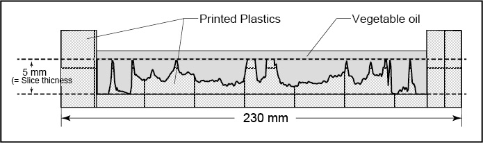
First, a transverse of spin echo T1WI in basal ganglia was acquired in a healthy subject as the model T1WI (female, 36 years) using 3.0T MRI unit (Achieva 3.0T-TX, Philips, the Netherlands) combined with a 32-channel SENSE head coil. Next, MATLAB R2015b (Mathworks Inc., Natick, MA, USA) was used to convert the DICOM data of the model T1WI to the STL file for 3D-printing in the following processes: (1) making profile curves in all line on the model T1WI (Fig. 2), (2) converting into thickness of the printed layer stack (T) by the following formula;T = (Imax – I) {(Tslice / Tlayer) / Imax} Tlayer + Tbottom
where Imax is the maximum signal intensity of the model T1WI, Tslice is the slice thickness of the T1WI, Tlayer is the thickness of a printed layer, and Tbottom is the thickness of the bottom of the phantom. Then, the profile curves were flipped vertical and the signal intensities were transformed to depths between 0 to 5 mm, which were converted to cross sections of the phantom (Fig. 3). Then, the converted data was sent to the 3D-printer (Objet Eden350V, Stratasys, USA), and printed by the plastic (FullCure810 Vero Clear), which has no MR signal. After printed, vegetable oil was poured onto the convexo-concave surface on the printed material. Finally, a spin echo T1WI of a single slice (5 mm thickness) that was planed to the convexo-concave of the phantom was acquired. The correlation coefficient between the model image and the phantom image was estimated using also MATLAB.
Results:
Figure 4 shows the outline of the phantom without vegetable oil. And, Figure 5 shows the T1WI of the model (a) and the phantom (b). We can see that the model in vivo T1WI and the T1WI of the phantom were in very good agreement. Furthermore, the correlation coefficient (R2) between the model image and the T1WI of the phantom was 0.941 (p < 0.001).Conclusions:
We finally could achieve to make the brain MRI phantom for T1- and T2-weighted image showing image contrast and construction similar to those of in vivo MRI by using 3D rapid prototyping technique. Our phantoms might be powerful tools to use for experiments that need ethically unacceptable long scanning or systematic movements: optimal parameters verification for the compressed sensing technique or understanding PROPELLER algorithm’s capabilities in real clinical situation.Acknowledgements
No acknowledgement found.References
1. Saotome K, Matsushita A, Matsumoto K, et al. A brain phantom for motion-corrected PROPELLER showing image contrast and construction similar to those of in vivo MRI. Magn Reson Imaging. 2017;36:32–39.
2. V. Filippou, C. Tsoumpas. Recent advances on the development of phantoms using 3D printing for imaging with CT, MRI, PET, SPECT, and ultrasound. Med. Phys. 2018;Sep.45(9):740-760.
Figures