S77
GRAPPA vs SMS Fast Brain MRI Compared to ACR Specifications
Mark J Burton1, Robert Thomen1, Talissa A. Altes1, Carlos Leiva Salinas1, Nicholas Tustison2, and Joseph P Cousins1
1Radiology, University of Missouri Hospital, Columbia, MO, United States, 2Radiology, University of Virginia, Charlottesville, VA, United States
1Radiology, University of Missouri Hospital, Columbia, MO, United States, 2Radiology, University of Virginia, Charlottesville, VA, United States
Synopsis
This work was to assess the trade-off between scan time and image quality afforded by acceleration techniques GRAPPA and simultaneous multi-slice (SMS). We imaged an ACR phantom using standard clinical T1, T2, and FLAIR sequences. Then, without altering any other scan parameters, we increased GRAPPA and SMS acceleration factors (2, 4, and 6). All GRAPPA and SMS accelerated scans passed the ACR resolution criteria. Both GRAPPA and SMS passed low-contrast criteria at acceleration factor 2, but factors of 4 and 6 did not. GRAPPA and SMS gave nearly identical similarity indices and SNR at all acceleration factors.
Purpose
In many institutions the standard brain MRI examination is a relatively lengthy imaging protocol. There is potential to substantially decrease acquisition time using recent technical developments including parallel imaging and simultaneous multi-slice [1-3]. However, these acceleration techniques can degrade image quality. The purpose of this work was to assess the trade-off between scan time and image quality using the industry standard ACR imaging quality metrics and the acceleration techniques GRAPPA and simultaneous multi-slice (SMS).Methods
MRI scans of the ACR detail phantom were acquired on a Siemens VIDA 3.0T MRI scanner (Siemens Healthcare, Erlangen, Germany) using a 20-channel head coil. T1-weighted, T2-weighted, and FLAIR images were acquired using the ACR standard for evaluating SNR and CNR high and low contrast object detectability measures. For each weighted sequence, all sequence parameters remained the same except the acceleration technique and acceleration factor (Table 1). Baseline scans were acquired without any acceleration techniques. GRAPPA and SMS were then added separately, and increased by 2, 4, and 6-times acceleration. The SNR, CNR, and minimum detectable element size were evaluated for each acquisition. The MR images were evaluated with spatial normalization, using quantitative comparison, and image similarity metrics [4], including localized neighborhood cross correlation [5] and the structural similarity index [6]. A visual evaluation of the high contrast resolution, low contrast and artifacts as set by the ACR, as minimum passing requirements, was also performed [7].Results
Figure 1 demonstrates the changes in SNR and similarity index for the T1, T2 and FLAIR acquisitions using GRAPPA and SMS acceleration factors of 2, 4 and 6. GRAPPA and SMS had nearly identical results in SNR and similarity indices for each acceleration factor. Both GRAPPA and SMS showed a slight increase in SNR at an acceleration factor of 2 but reduction in SNR and similarity indices at acceleration factors of 4 and 6. All GRAPPA and SMS accelerated scans passed the ACR resolution and high-contrast criteria, but only the acceleration factor 2 scans passed the ACR low-contrast criteria (Figure 2 and 3).Conclusions
By assessing resolution, high- and low-contrasts, SNR, and similarity indices between images of increasing acceleration, we find that GRAPPA and SMS acceleration factor of 2 gave the best imaging results with increased SNR while reducing the scan time by 40%, but higher acceleration factors of 4 and 6 dramatically reduced SNR and produced images of non-diagnostic low-contrast quality according to ACR quality assurance guidelines.Acknowledgements
No acknowledgement found.References
1. Wiggins GC, Polimeni JR, Potthast A, et al. 96-channel receive-only head coil for 3 Tesla: design optimization and evaluation. Magn Reson Med 2009;62:754 –62 2. Wiggins GC, Triantafyllou C, Potthast A, et al. 32-channel 3 Tesla receive-only phased-array head coilwith soccer-ball element geometry. Magn Reson Med 2006;56:216–23 3. Keil B, Blau JN, Biber S, et al. A 64-channel 3T array coil for accelerated brain MRI. Magn Reson Med 2013;70:248 –58 4. Avants BB, Tustison NJ, Song G, Cook PA, Klein A, Gee JC. A reproducible evaluation of ANTs similarity metric performance in brain image registration. NeuroImage. 2011;54(3):2033-44. 5. The Insight ToolKit image registration framework. Avants BB, Tustison NJ, Stauffer M, et al. Front Neuroinform. 2014;8:44. 6. Image quality assessment: from error visibility to structural similarity. Wang Z, Bovik AC, Sheikh HR, Simoncelli EP. IEEE Trans Image Process. 2004;13:600-12. 7. American College of Radiology MR Accreditation Program Testing Instructions. (accessed Nov 6, 2019) https://www.acraccreditation.org/-/media/ACRAccreditation/Documents/MRI/MRAccreditationTestingInstructions.pdf?la=enFigures
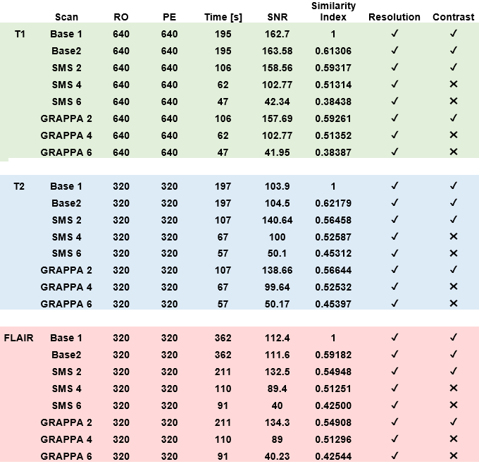
Table 1: Imaging Parameters: Scan time, SNR, and Similarity Indices, and
a pass/fail (✓,
✖) analysis for ACR resolution and
contrast criteria [7].
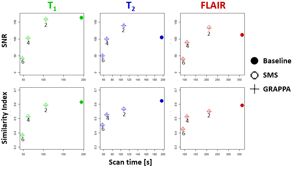
Figure 2. Comparison of SMS to GRAPPA techniques with accelerations factors 2, 4, and
6 in sagittal T1, axial T2 and axial FLAIR imaging, compared to standard
acquisition (baseline).
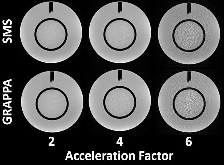
Figure
2. Example T2 images of the ACR phantom comparing SMS and GRAPPA techniques
with accelerations of 2, 4, and 6. Notice both acceleration techniques “pass”
the ACR requirements at acceleration factor of 2, while loss of CNR and
additional noise lead to “failed” the scans at acceleration factors of 4 and 6.