S76
What Information Can Diffusion Tensor Fractional Anisotropy Provide Relative to Environmental Enrichment in Children with Congenital Heart Defects?1Radiology, University of Pittsburgh Medical Center, Pittsburgh, PA, United States, 2Critical Care Medicine, University of Pittsburgh Medical Center, Pittsburgh, PA, United States, 3Behavioral Health Sciences, University of Pittsburgh Medical Center, Pittsburgh, PA, United States, 4Physical and Occupational Therapies, University of Pittsburgh Medical Center, Pittsburgh, PA, United States
Synopsis
Children with Congenital Heart Defects (CHD) are at an increased risk for neurological deficits and neurodevelopmental delays with the most common anomaly being white matter injury (WMI). Diffusion tensor imaging (DTI), uses water’s diffusivity to characterize microstructural changes in WM. One DTI metric, fractional anisotropy (FA), increases with myelination and decreases in the presence of demyelination. We examined the association between early developmental therapies (such as OT, PT, and SLP) and WMI in infants with CHD using FA as a quantitative imaging metric for white matter integrity.
Background
It is well-established that children with Congenital Heart Defects (CHD) are at an increased risk for neurological deficits and neurodevelopmental delays1. The most common neurological anomaly in infants with CHD is white matter injury (WMI). Diffusion tensor imaging (DTI), uses water’s diffusivity to characterize microstructural changes in WM. One DTI metric, fractional anisotropy (FA), increases with myelination and decreases in the presence of demyelination2. Increasing evidence is highlighting the importance of environmental impact on myelination. Many pediatric cardiac intensive care units use family-based, multidisciplinary developmental care strategies to “enrich” early environment and improve neurodevelopmental outcomes. We examined the association between early developmental therapies and WMI in infants with CHD using FA as a quantitative imaging metric for white matter integrity.Methods
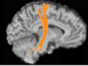
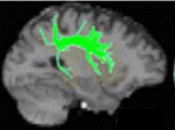
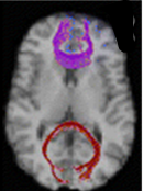
16 post-operative MRI scans (PCA < 4 months) were obtained on a 3T MAGNETOMTM Skyra (Siemens, Erlangen, Germany) using the 32-channel head-array coil (5 infants also had a pre-operative MRI used for FA trajectory.) 42 direction DTI with B = 1000s/ mm2, TE/TR=92ms/12600ms, and 2.0mm isometric resolution were acquired. Eddy current correction was performed followed by tractography using DSI studio to calculate FA. Tracts examined: left (L) and right (R) tracts of cortico-spinal tract (CST) (Figure 1), fronto-occipital fasciculus (FOF) (Figure 2), superior and inferior longitudinal fasciculi (SLF and ILF; anterior (A) and posterior (P)) (Figures 3 and 4), and genu, body, and splenium of the corpus callosum (CC) (Figure 5). Retrospective review of the infant’s medical record was used to gather developmental care measures: length of stay (LOS); presence of occupational (OT), physical (PT), speech (SLP), and music (MT) therapies; number of therapy sessions by discipline, success of therapy by discipline, infant mobility, and family presence. FA and developmental care variables were compared using multiple linear and logistical regression with post-conceptual age at scan as a covariate.Results
Family participation positively associated with FA within the ILF-R (p=0.0183). Infant mobilization also positively associated with FA trajectory within the SLF-R and SLFP-R (p=0.029 & 0.038, respectively). Success rates for OT were positively associated with FA within the CST-R (p=0.014), ILF-L (p=0.048), SLF-R (p=0.024), and SLFA-L (p=0.046) while speech success correlated with FA elevations within the CST-R (p=0.024) and FOF-R (p=0.03). In contrast, FA was negatively associated with PT consult in the CC (P=0.042), CST-L (P=0.023), Genu (p=0.01); while OT consult negatively associated with FA in the SLF-R (p=0.035) (Table 1). LOS, MT consult, number therapeutic sessions per discipline (PT, OT, SLP, and MT), and days to feeding failed to show any significant association with FA in any of the analyzed brain regions.Conclusion
Together, our data suggest that family presence and infant mobility may support region-specific WM myelination in infants with CHD. OT and SLP success rates were also associated with increased myelination. However, the number of sessions showed decreased myelination, which may indicate that an infant’s post-operative status may outweigh other extrinsic factors in prognosticating neurological deficit. We continue to examine this relationship in a larger sample size and how these metrics and variables impact functional outcomes.Acknowledgements
Christine Johnson - Pediatric Imaging Research Center; the Clinical MRI Staff at UPMC Children’s Hospital of Pittsburgh - Department of RadiologyReferences
1 Nattel SN, Adrianzen L, et al. Congenital Heart Disease and Neurodevelopment: Clinical Manifestations, Mechanisms, and Implications. Canadian Journal of Cardiology; 2017; 33, 1543-1555
2 Schmithorst VJ, Votava-Smith JK, et al. Structural network topo correlates of microstructural brain dysmaturation in term infants with CHD. Human Brain Mapping 2018; 39, 4593-4610
Figures