Metabolite concentrations in the parietal white matter: Is there a difference between left and right hemispheres in young children?
1Cape Universities Body Imaging Centre, University of Cape Town, Cape Town, South Africa, 2Department of Human Biology, Faculty of Health Sciences, University of Cape Town, Cape Town, South Africa, University of Cape Town, Cape Town, South Africa, 3Department of Paediatrics and Child Health, Red Cross War Memorial Children’s Hospital, University of Cape Town, SA, University of Cape Town, Cape Town, South Africa, 4Neuroscience Institute, University of Cape Town, Cape Town, South Africa, 5Department of Clinical Research, London School of Hygiene & Tropical Medicine,, London, United Kingdom
Synopsis
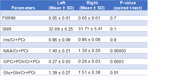
We retrospectively analyzed 1HMRS data from right and left parietal white matter in a study of 2-year-old children, aiming to determine whether there was a difference between right and left hemispheres in metabolite ratios to creatine. We found concentration ratios of NAA and choline to be higher on the left and Glu+Gln higher on the right. Correlation between right and left was highest for myo-inositol (r= 0.81), but fairly low for NAA (r = 0.63) and Glu+Gln (r = 0.42). Consideration should be given to hemisphere selection when planning 1HMRS studies in young children.
Background
1HMRS measures local brain metabolism, providing information about biochemical aspects of brain maturation. The metabolites typically measured with 1HMRS include creatine (Cr+PCr), N-acetyl-aspartate (NAA), choline (GPC+PCh) and glutamate (Glu), which are associated with energy metabolism, neuronal and cellular integrity and neurotransmission. These metabolites have been implicated in ongoing myelination in a longitudinal study of five to 10-year-old children examining regional age-related changes in metabolite levels. In particular, increased levels of Cr+PCr, NAA, NAA+N-acetyl-aspartyl-glutamate (NAA+NAAG), Glu and combined glutamate+glutamine (Glu+Gln) were found in white matter with age1.Rationale
Part way through collecting MRS data from parietal white matter in high risk 2-year old children from a South African birth cohort, we noticed an inconsistency in the hemisphere from which 1HMRS data were being collected. This prompted additional data collection from both right and left parietal white matter regions in subsequent participants for maximum consistency and in order to understand any differences between hemispheres. Clinical data provide strong evidence for hemispheric functional asymmetry, supporting the potential for metabolite concentrations to differ between hemispheres2. However, metabolite ratios in adults have been found to be symmetric in most brain regions, except the thalamus3, and in a study on children from 6 – 16 no hemispheric asymmetries in metabolite ratios to creatine were found4.Objectives
The primary objective of this pilot study was to determine whether there is a difference in metabolite concentrations in the right and left parietal white matter regions in developing children. This data will determine whether spectra from both hemispheres could be included in the data analysis for the main study. We hope that this pilot study can be used as a reference to guide future studies in selecting whether to collect single voxel spectroscopy from one or both hemispheres.Methods
We retrospectively evaluated the MRS data obtained from 59 healthy, 2-year-old children, 29 males and 30 females, from the Drakenstein Child Health study5. All scans were done on a 3T Magnetom Skyra, Siemens (Erlangen Germany), scanner using a 32-channel head coil. Spectroscopy of the 1H nucleus was performed using a point-resolved (PRESS ) single voxel MRS sequence with TR 2000ms; TE 30ms; averages 128; spectral bandwidth 1200Hz ; vector size 1024 and 2.5x2.5x2.5 cm voxel size. Voxel placement was in the right and left parietal white matter regions. Automatic shimming was performed before acquiring the MRS. The mean linewidth recorded by the scanner was 14.2 MHz and 14.1 MHz for the left and right hemispheres respectively. The ratios to creatine of four metabolites , namely choline, N-acetylaspartate, myo-inositol, and glutamate/glutamine complex (Glx), were estimated using LCModel. Data with FWHM>0.08 ppm reported by LCModel were excluded. Systematic differences in metabolite to creatine ratios were investigated using paired t-tests. Pearson correlation was performed to assess the correspondence between right and left hemisphere metabolites across subjects.Results
Paired t-tests showed that concentrations of NAA and choline are respectively ~8% and ~4% higher on the left (p’s<0.005) and Glu+Gln ~9% higher on the right (p<0.005) (see Table 1). Correlation between right and left is highest for myo-inositol (r= 0.81), but fairly low for NAA (r = 0.63) and Glu+Gln (r = 0.42) (see Figure 1).Conclusion
Despite previous findings of no metabolite ratio asymmetries in older children, we observe differences in concentrations of NAA, choline, Glu+Gln ratios between right and left hemispheres in a healthy cohort of 2-year-old children. Certain subjects show quite large asymmetries in NAA and Glu+Gln (fig.1). Therefore, studies examining metabolite group differences in developmental disorders cannot be certain that findings would be the same for right and left hemispheres, particularly for NAA and Glu+Gln. These findings are useful to consider when analyzing the data for the main study.Acknowledgements
Mazwi Maishi
Ingrid Opt'Hoff
Judy Gatei
Annerine Roos
Joavine Fourie
Tabitha Mutseyekwa
Sivenesi Subramoney
Katherine L. Narr
Dan J Stein
Heather J Zar
References
1. Holmes, MJ., Robertson, FC., Little, F., Randall, SR., Cotton, MF., Van der Kouwe, AJW., Laughton, B., Meintjes, EM. Longitudinal increases of brain metabolite levels in 5-10-year-old children: PLOS ONE | https://doi.org/10.1371/journal.pone.0180973
2. Toga AW , Thompson PM. 2003. Mapping brain asymmetry. Nat Rev Neurosci . 4:37–48
3. Nagae-Poetscher LM1, Bonekamp D, Barker PB, Brant LJ, Kaufmann WE, Horská A. Asymmetry and gender effect in functionally lateralized cortical regions: a proton MRS imaging study: J Magn Reson Imaging. 2004;19(1):27–33. PubMed Google Scholar
4. Cichocka, M.; Kozub, J.; Karcz, P.; Urbanik, A. Differences in Metabolite Concentrations Between the Hemispheres of the Brain in Healthy Children: A Proton Magnetic Resonance Spectroscopy Study (1HMRS): Journal of Child Neurology 2016, Vol. 31(11) 1296-1301 https://doi.org/10.1177/0883073816653784
5. Donald, KA.; Hoogenhout, M.; du Plooy, CP.; Wedderburn, CJ.: Nhapi, RT.; Barnett, W.; Hoffman, N.; Malcolm-Smith, S.; Zar, HJ, and Stein, DJ. Drakenstein Child Health Study (DCHS): investigating determinants of early child development and cognition: BMJ Paediatrics Open 2018;2:e000282. https://doi:10.1136/ bmjpo-2018-000282
Figures