S59
What is the appropriate number of acceleration factor of compressed sensing with diffusion weighted image?1Division of Radiology and Nuclear Medicine, Sapporo Medical University Hospital, Sapporo, Japan
Synopsis
In several recent studies, the compressed sensing (CS) technique has been applied to various sequence, which helped reduce the scan time while showing minimal effect on image quality. However, the appropriate number of acceleration factor of CS is unclear. We aimed to reveal the appropriate number of acceleration factor of CS with TSE-DWI. We calculated %CV from the image of ADC, the similarity between images of the control and the accelerated sequences was evaluated using structural similarity index (SSIM). From results of this research, the appropriate number of acceleration factor of CS with TSE-DWI was revealed as up to 4.
Background and Purpose
The compressed sensing (CS) technique is a method to accelerate MR acquisition by acquiring undersampling k-space data. In several recent studies, the CS technique has been applied to various sequence of MRI, which helped reduce the scan time while showing minimal effect on image quality [1]. Whereas the turbo spin echo (TSE) technique is an alternate approach for diffusion-weighted imaging (DWI). Because it uses a 180° RF refocusing pulse for each measured echo, susceptibility artifacts and image distortion are lower than on echo-planar imaging DWI scans. Previous reports suggested that decrease in the SNR and a long acquisition time are disadvantages of TSE-DWI [2]. Thus, TSE-DWI combined with CS may be useful to short acquisition time with keeping SNR, however, the appropriate number of acceleration factor of CS is unclear. The aim of this study was to analyze the change of image quality and ADC with increasing of acceleration factor, and reveal the appropriate number of acceleration factor of CS with TSE-DWI.Methods
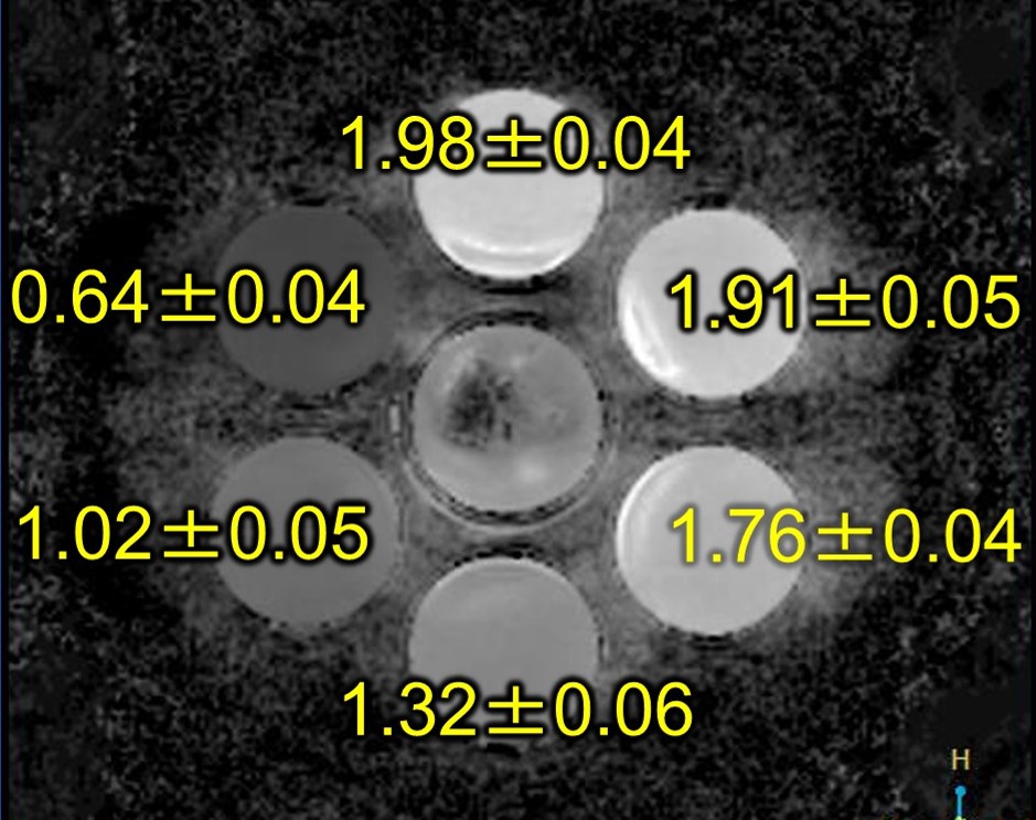
We performed using a 3-Tesla MR system (Ingenia, Philips Healthcare), and created homemade phantom with changed concentration of polyethylene glycol (PEG) to obtain different ADC (Fig. 1). The basic scan technique of TSE-DWI was set at TR, 5000ms; TE, 75ms; ETL, 128; b value, 1000. The image obtained from this basic technique was used as a control image. Acceleration factor of CS was changed from 2 to 20. We calculated %CV from image of b0, b1000 and ADC. The similarity between images of the control and the accelerated sequences was evaluated using structural similarity index (SSIM).Results
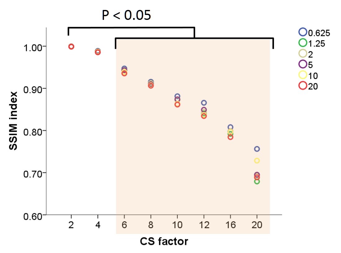
The %CV of b0, b1000 and ADC with control image were 2.42 ± 0.82, 5.34 ± 0.21 and 0.49 ± 0.25, respectively. Acceleration factor more than 8 had significantly higher %CV of ADC (p < 0.05) (Fig. 2). Whereas the mean value of SSIM index with acceleration factor from 2 to 20 was 0.999 to 0.707. The SSIM showed significant decrease with acceleration factor more than 6 (Fig. 3).Conclusion
The appropriate number of acceleration factor of CS with TSE-DWI was up to 4.Acknowledgements
No acknowledgement found.References
1. R. Kijowski, H. Rosas, A. Samsonov, et al. Knee imaging: rapid three-dimensional fast spin-echo using compressed sensing, J. Magn. Reson. Imaging 45; 1712–1722, 2017.
2. CJ Juan, HC Chang, CJ Hsueh, et al. Salivary glands: echo-planar versus PROPELLER Diffusion-weighted MR imaging for assessment of ADCs. Radiology 253:144–52, 2009.
Figures