S33
Effectiveness of distortion correction for diffusion-weighted imaging using clinical images1Tokushima Bunri University, Sanuki, Japan, 2Division of Health Sciences, Graduate School of Medical Sciences, Kanazawa University, Kanazawa, Japan, 3Kyoto University Hospital, Kyoto, Japan, 4Otsu City Hospital, Otsu, Japan, 5The University of Tokyo Hospital, Bunkyo-ku, Japan
Synopsis
We investigated whether our distortion correction method could be applied practically for clinical breast MRI. The cross-correlation coefficients (CCCs) of the whole image and high-intensity regions (HIR) were calculated to assess the correlations between b0 and FS-T2WI images and b1500 and T1CE images. For all these combinations of images, the CCCs showed higher correlations with distortion correction than without the correction. This method could be adapted to any type of breast and HIR. Our method is particularly helpful as there is no additional scan and no extension in the scan time.
Background
Diffusion-weighted images (DWIs) are widely used for tumor differentiation or for assessing treatment effects and are considered to be one of the essential sequences for breast magnetic resonance imaging (MRI). We therefore developed a retrospective distortion correction technique for breast MRI based on non-rigid image registration, without the need for extra scanning [1]. A phantom study showed that compared with TOPUP [2], a previous study resulted in more accurate cross-correlation coefficients (CCCs), shape-error analysis, and ADCs [1].Purpose
The aim of the present study was to investigate whether our distortion correction method could be applied practically for clinical breast MRI.Methods
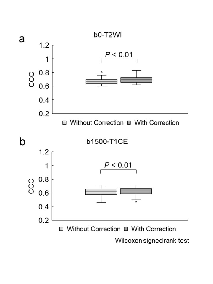
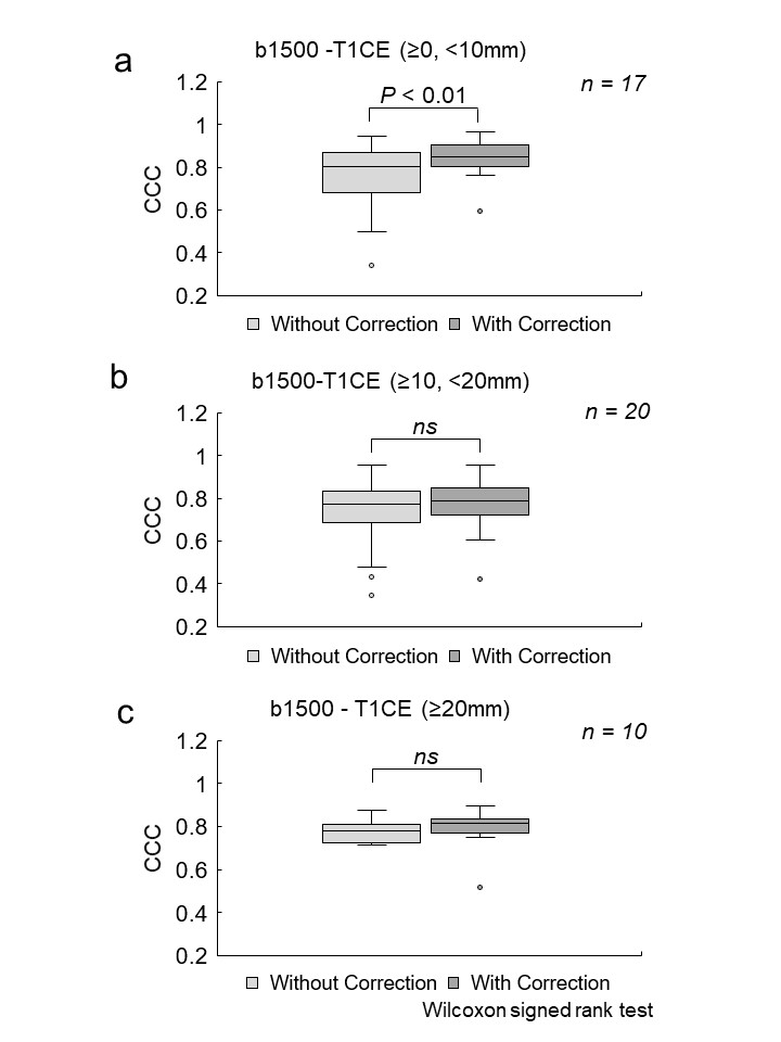
A 3.0T MRI scanner with an 18-channel dedicated breast coil was used. The diffusion-weighted imaging was used for scan. The proposed study included several steps, which are FOV size matching, matrix size matching, image segmentation, edge detection, non-rigid image registration, and image wrap. We applied the method to data for 50 consecutive breast cancer patients (age, 56.9 ± 13.0 years), evaluating it for whole images and for various sizes of high-intensity regions (HIR). To assess the distortion correction of EPI for the contour of the entire breast, cross-correlation coefficients (CCCs) of the whole image and HIR were calculated to assess the correlations between b0 and FS-T2WI images and b1500 and T1CE images. The CCCs were calculated for comparing the uncorrected and corrected images, with higher values indicating a greater correlation. Wilcoxon signed rank tests were used for the statistical analysis of the comparisons. The mean (± standard deviation) long and short diameters of the ROIs for the 47 HIRs were 15.0 ± 10.5 mm and 10.0 ± 7.1 mm, respectively. To evaluate whether the distortion correction was affected by the size of the HIR, percentage differences in CCCs with and without correction were calculated, depending on sizes of HIR, on the basis of the long diameter of the ROI. The median (IQR) CCCs were calculated for HIRs of different sizes [“≥0, <10” mm (n = 17), “≥10, <20”mm (n = 20), and “≥20”mm (n = 10)] with and without distortion correction, and the mean percentage differences between them were evaluated and analyzed using the Wilcoxon signed rank test.Results
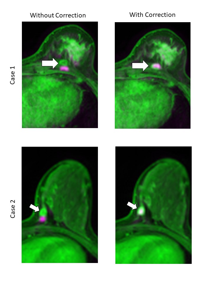
For all these combinations of images, the CCCs showed higher correlations with distortion correction than without the correction. All the differences were significant (P < 0.01) (Fig. 1). The size “≥0, <10” mm: indicating a significant difference (P < 0.01); size “≥10, <20”mm: indicating a non-significant difference (P = 0.064); and size “≥20”mm: indicating a non-significant difference (P = 0.084) (Fig. 2). The distortion correction effect was relatively small when the HIR was large. Sample images are presented in Fig. 3.Conclusions
Our distortion correction method can be applied to clinical breast MRI. This method could be adapted to any type of breast and HIR, especially small region. Our method is particularly helpful as there is no additional scan and no extension in the scan time.Acknowledgements
Nothing in particular.References
[1] Takatsu Y, et al. Novel distortion correction method for diffusion-weighted imaging based on non-rigid image registration between low b value image and anatomical image. Magn Reson Imaging 2019; 57:277–284.
[2]Wan X, et al. Reduction of geometric and intensity distortions in echo-planar imaging using a multireference scan. Magn Reson Med., 1997;37(6):932–942.
Figures