S32
Useful sequence for suppressing the radiofrequency shielding effect of a titanium mesh1Tokushima Bunri University, Sanuki, Japan, 2Division of Health Sciences, Graduate School of Medical Sciences, Kanazawa University, Kanazawa, Japan, 3Kurihara Central Hospital, Kurihara, Japan, 4Osaka Medical College Hospital, Takatsuki, Japan, 5Nara Medical University Hospital, Kashihara, Japan
Synopsis
To determine which sequence for routine contrast-enhanced brain MRI (2-dimentional spin echo, 2D SE; and 3-dimentional gradient echo, 3D GRE) exhibits less RF shielding effect of the titanium mesh after cranioplasty, using a phantom in multi vendor. Every vendor had similar signal attenuation ratio and normalized absolute average deviation (NAAD) in the proximal section of phantom image. With regard to NAAD, there was a significant difference between 2D SE and 3D GRE (P < 0.05). 3D GRE shows less RF shielding effect of a titanium mesh, and it might be appropriate sequence for MRI when a titanium mesh is present.
Background
One of the most commonly used materials for cranioplasty is a titanium mesh owing to its excellent biocompatibility and mechanical performance [1]. However, a titanium mesh has been reported to cause radiofrequency (RF) shielding [2]. After surgery for brain tumors, T1-weighted imaging (T1WI) with contrast medium is performed to assess the presence of remnant tumors and recurrence [3]. However, the influence of the RF shielding effect with the use of a titanium mesh is a concern. If the RF shielding effect is reduced using suitable routine sequences, high-quality images will be obtained.Purpose
The present study aimed to determine which sequence for routine contrast-enhanced brain MRI (2-dimentional spin echo, 2D SE; and 3-dimentional gradient echo, 3D GRE) exhibits less RF shielding effect of the titanium mesh after cranioplasty, using a phantom.Methods
A 1.5-T MRI scanners (Philips, Achieva; Siemens, MAGNETOM Avanto; and GE Healthcare, Signa Explorer) were used for imaging. The sequence parameters were as uniform as possible. A cubic bottle [152 × 152 × 195 mm (width × height × depth)] filled with 33 mg/L of manganese chloride tetrahydrate and 3.6 g/L of sodium chloride was used as a phantom to mimic the human brain. The phantom was placed at the center of the coil, and a titanium mesh board (100 × 100 × 0.8 mm; hole diameter, 1.6 mm; hole interval, 3 mm; thickness, 0.8 mm), which is identical to the material used during actual cranioplasty, was placed on the phantom. The signal attenuation ratio was calculated as the signal intensity of the image obtained in the presence of the titanium mesh divided by the signal intensity obtained in the absence of the mesh. If the signal intensity did not change, the signal attenuation ratio was 1 [2]. To evaluate uniformity as RF shielding dependency, normalized absolute average deviation (NAAD) was used. NAAD was calculated using the following equation:$$NAAD=100\times[1-\frac{1}{N\bar{Y}}\sum_{i=1}^N(\mid Yi-\bar{Y}\mid)]$$
where$$$Yi$$$is the individual pixel value within the measurement region of interest (MROI), $$$\bar{Y}$$$is the mean of all pixels within the MROI,$$$\mid Yi-\bar{Y}\mid$$$is the absolute deviation for pixel $$$i$$$, and $$$N$$$ is the total number of pixels within the MROI.
Conventional 2D SE and 3D GRE were compared in the proximal section of the titanium mesh (most influenced by RF shielding). The Wilcoxon paired-rank test were used for statistical analyses (EZR v. 3.4.1). A P-value <0.05 was considered statistically significant.
Results
3D GRE showed less radiofrequency shielding effect than 2D SE, and it was considered as the optimal sequence for MRI in the presence of a titanium mesh. Every vendor had a similar signal attenuation ratio (Fig. 1) and NAAD in the proximal section (Fig. 2). With regard to NAAD, there was a significant difference between 2D SE and 3D GRE (P < 0.05) (Fig. 2). Fig. 3 presents sample images with contrast medium.Conclusions
3D GRE shows less RF shielding effect of a titanium mesh after cranioplasty among routine sequences for contrast-enhanced brain MRI, and it might be the most appropriate sequence for MRI when a titanium mesh is present.Acknowledgements
Nothing in particular.References
[1] Cabraja M, et al. Long-term results following titanium cranioplasty of large skull defects. Neurosurg Focus 2009; 26:1–7.
[2] Takatsu Y, et al. Radiofrequency-shielding Effect of a Titanium Mesh Implanted for Cranioplasty. Magn Reson Med Sci 2015;14(4):321-327.
[3] Niendorf HP, et al. Dose administration of gadolinium-DTPA in MR imaging of intracranial tumors. Am J Neuroradiol 1987;8(5):803–815.
Figures
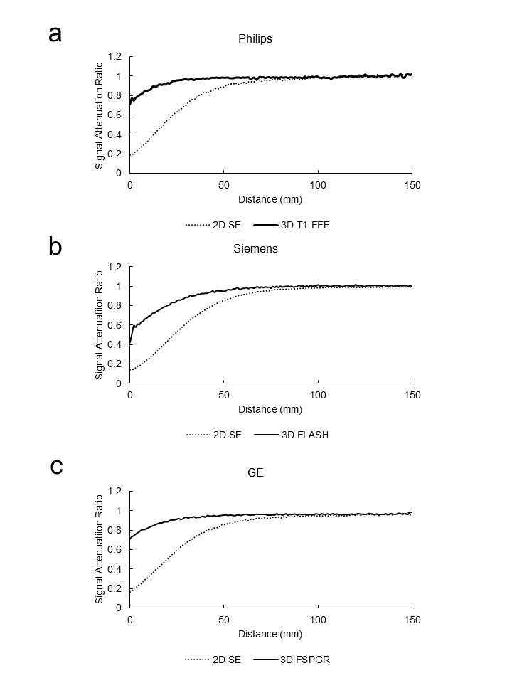
Fig. 1; Signal attenuation ratio of 2D SE and 3D GRE.
Philips (a), Siemens (b), GE (c)
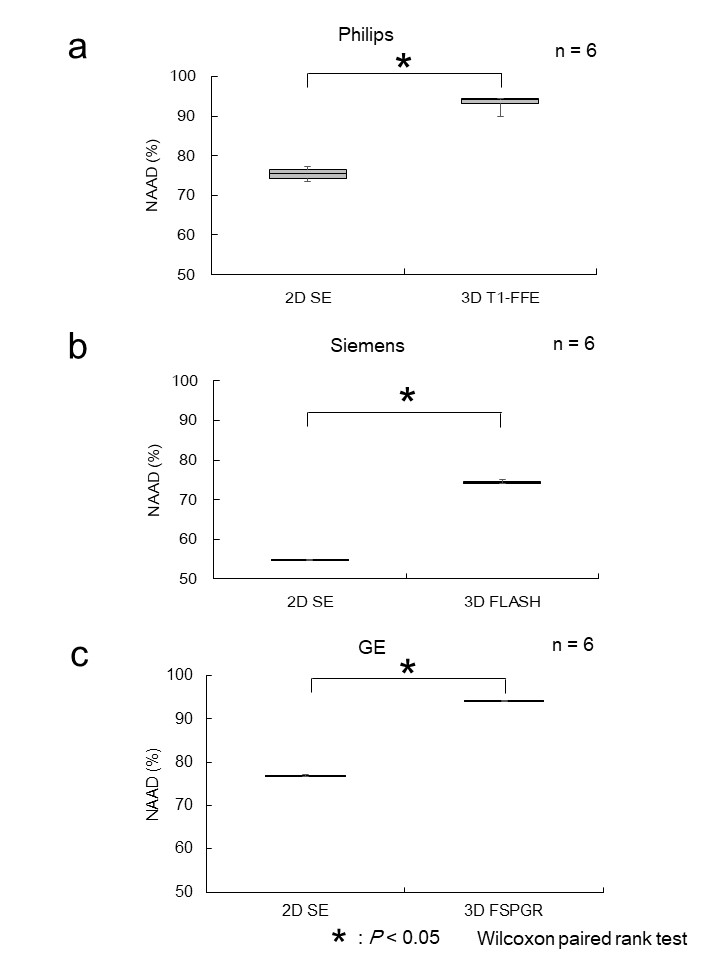
Fig. 2; NAAD in proximal area of phantom image.
2D SE and 3D GRE. Philips (a), Siemens (b), GE (c)
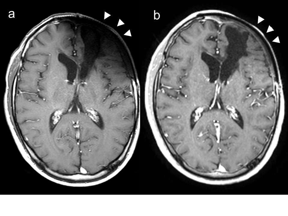
Fig. 3; Sample images of 2D SE (a) and 3D GRE (b)
Vicinity of titanium mesh (arrow head) of 3D GRE was clearer visualized than 2D SE.