S31
Performance of Automatic B0 Shim Modes for Brain MR Spectroscopy on 7T and 3T1Radiology, Brigham and Women's Hospital, Boston, MA, United States
Synopsis
For magnetic resonance spectroscopy, automated B0 shim mode may save scan time in a fast-paced clinical environment but is strongly dependent on performance. Two shim modes were compared in multiple brain regions at Siemens 3T and 7T. B0 maps were used to evaluate the shimming performance of each mode in the left hippocampus region. The results show that advanced shim mode is preferred on 7T (syngo MR VE12) and brain shim mode is preferred on 3T (syngo MR VE11) for brain MRS. However, more sophisticated shimming methods are still suggested for scanning more inhomogeneous regions if time permits.
Introduction
B0 shimming is particularly important for brain MR spectroscopy. A good shim with narrow spectral lines will provide more reliable separation and quantification of metabolites which in turn improves diagnostic accuracy1,2. Siemens MR software such as Syngo MR VE11 and VE12 provided different automatic B0 shim mode options that can save scan time by automatically optimizing the B0 field within the MRS voxel. This avoids time-consuming manual shimming which is not possible in a routine clinical environment. However, to our knowledge, the performance of these shim modes for brain MRS scan has not been tested. The goal of this study is to evaluate the shim modes’ performance and determine the ideal shim mode to use for brain MRS scan in clinically-relevant scenarios using both linewidth and B0 mapping comparisons.Methods
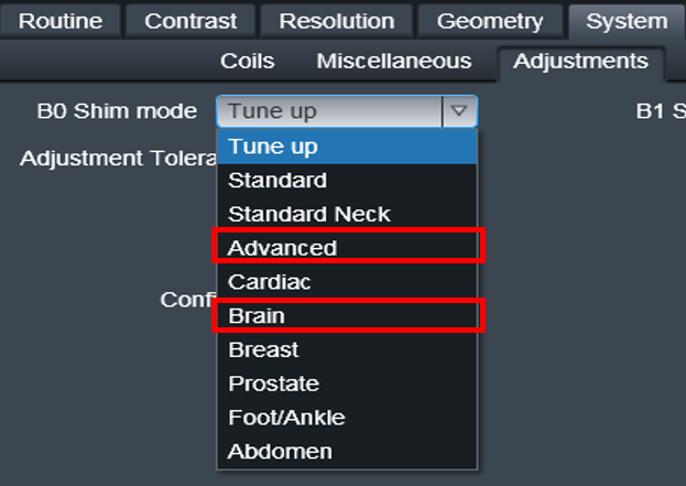
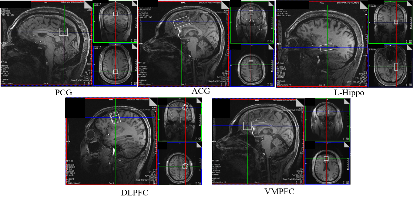
MR exams were performed in 14 subjects after informed consent on a Siemens Magnetom Terra 7 Tesla MRI scanner (Syngo MR VE12) using a Nova medical head coil (1TX / 32RX) with dielectric pads6 and on a Siemens Magnetom Skyra 3T MRI scanner (Syngo MR VE11) using 32 and 20 channel head coils.Full Width Half Maximum (FWHM), the spectral linewidth reported by Siemens Syngo system, were compared in different MRS voxel locations for two common B0 shim modes: “Brain” and “Advanced” (Figure 1) at 7T and 3T. Locations included: anterior cingulate gyrus (ACG), posterior cingulate gyrus (PCG), left hippocampus (L-hippo), dorsolateral prefrontal cortex (DLPFC), ventromedial prefrontal cortex (VMPFC) as shown in Figure 2. These regions were selected as they are often used across many different studies such as psychiatric diseases such as schizophrenia1,4 and bipolar 1,5 disorder. A focus on the left hippocampus was made because it was a region close to sinus with more field distortion and difficult to shim. At 3T, the comparison was also done in patient with a tumor adjacent to the right posterior ventricle. Paired t-tests was performed to compare the FWHM from the two shim modes.
3D B0 Mapping: of the left hippocampus region (3.5x1.5x1.5cm voxel) using the following parameters: TR/TE1/TE2=4.4/1.02/3.06ms, flip angle 10°, 44 slices and image acquisition time of 8.3s. As the adjust volume (shim box) was copied from the MRS voxel, B0 was set to the same shim channel values on X, Y, Z, Z², ZX, ZY, X²-Z² and XY as the MRS voxel in advanced shim mode. This was then repeated for brain shim mode. B0 maps were processed by MATLAB.
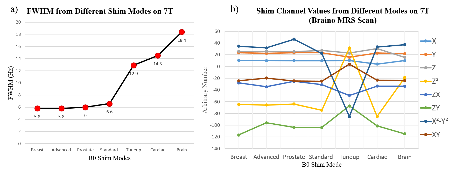
Additional B0 Shim Modes: A braino phantom with a 3x3x3 cm3 voxel was scanned with different shim modes to evaluate how shim channels being optimized on 7T. Shim modes included: Tune up, Standard, Advanced, Cardiac, Brain, Breast, and Prostate (Figure 1).
Results
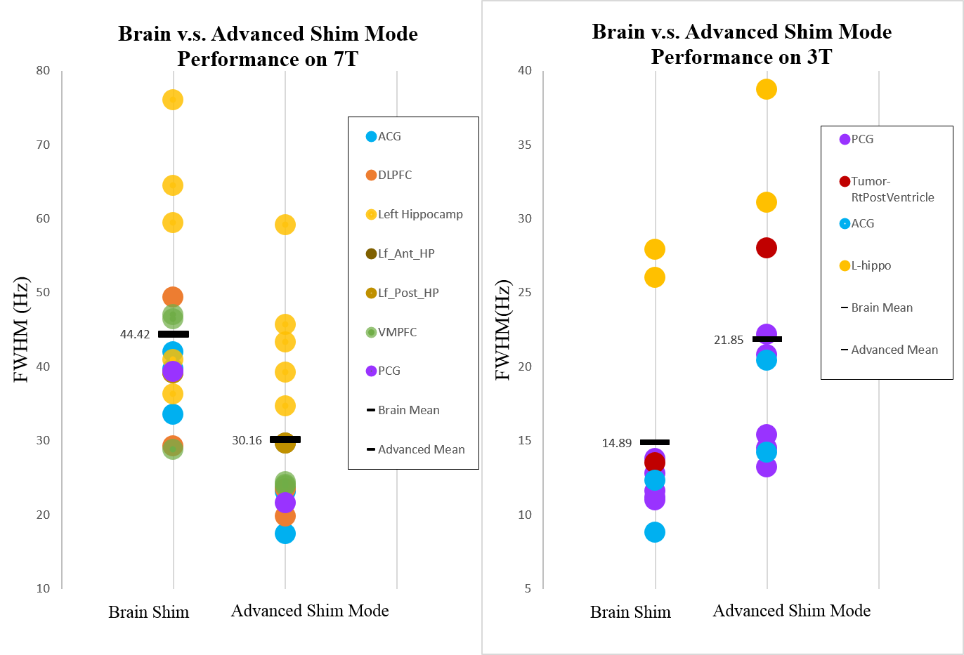
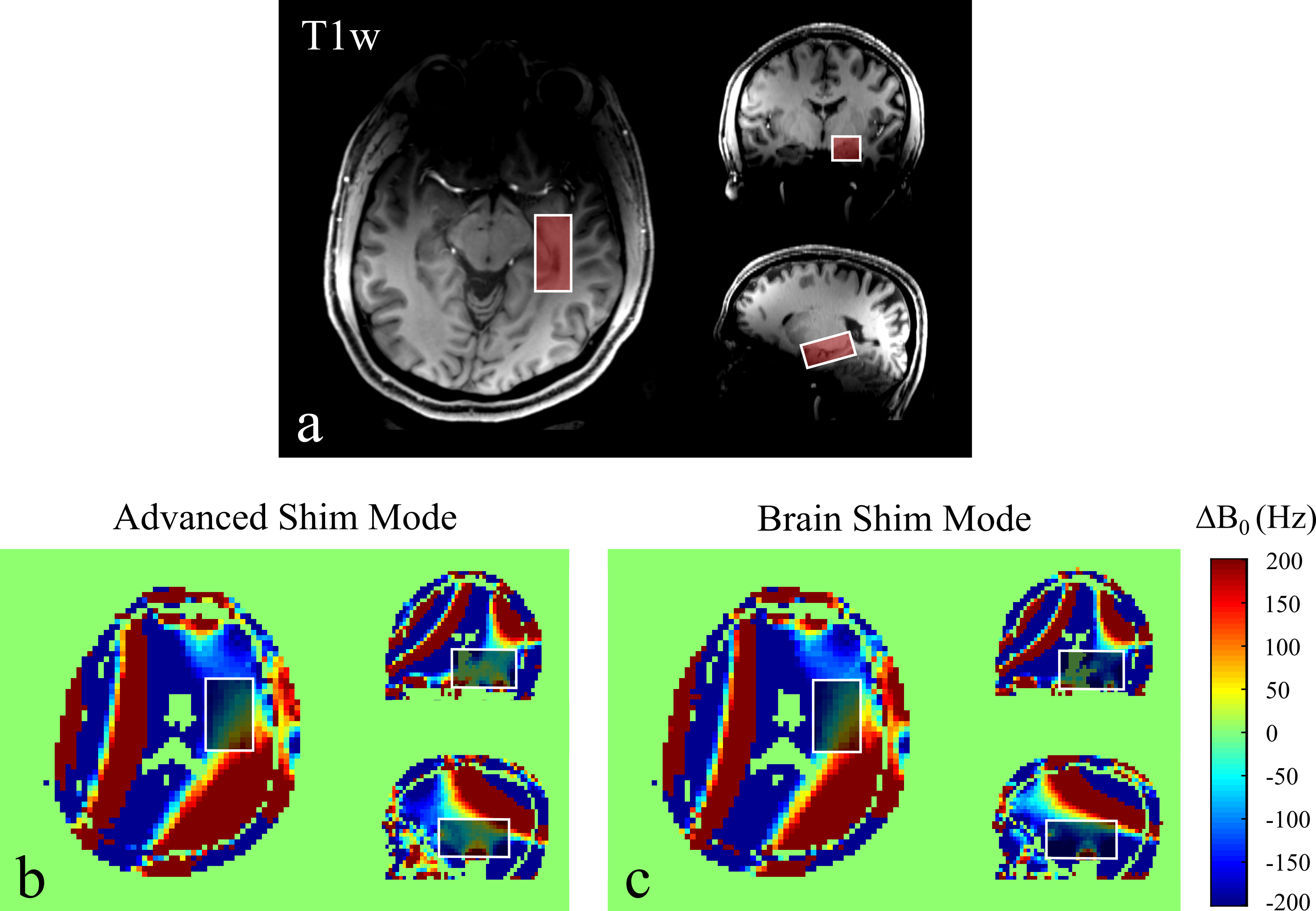
FWHM: Figure 3 shows a wide range of FWHM from different MRS voxel locations with higher FWHM in left-hippo (yellow) and lower FWHM in PCG (purple) and ACG (blue) in both shim modes as expected. However, brain shim mode (mean FWHM 14.89±6.53Hz) performs significantly better than advanced shim mode at 3T (mean FWHM 21.85±8.43Hz; p=0.0016), but at 7T, brain shim mode (mean FWHM 44.42±12.76Hz) performs significantly worse than advanced shim mode (mean 30.16±11.40Hz; p=0.00038).3D B0 Mapping: In a challenging region like the left hippocampus, brain and advanced shim mode give similar FWHM values (Brain shim:44.8Hz and Advanced shim:44.1Hz) on 7T. However, even with similar FWHM values, B0 maps show that advanced shim mode can generate a more homogeneous region (mean ΔB0 =111Hz) as the selected volume included the left hippocampus than brain shim mode (mean ΔB0 =141Hz) which is shown in Figure 4b and c (especially on the coronal and sagittal planes). However, when a smaller volume (the size of the left hippocampus voxel) is selected, both modes give the same mean ΔB0=80Hz.
Additional B0 Shim Modes: Figure 5a shows FWHM values rank from low to high (left to right) from different B0 shim modes. Interestingly, both breast and advanced shim modes give the best FWHM but brain gives the worst. FWHM from breast, advanced, prostate and standard shim modes are similar (5.8-6.6Hz) since they give similar values in first-order (X, Y, Z) channel (Figure 5b), but tune up, cardiac and brain shim modes give double even triple of FWHM since channel Y of tune up, X of cardiac and Z of brain give much lower values than other shim modes (Figure 5b). Also, all second order channels (Z², ZX, ZY, X²-Z², XY) behaved completely differently than others.
Discussion and Conclusion
The results show that advanced shim mode is preferred on 7T and brain shim mode is preferred on 3T for brain MRS exams. By choosing the correct shim mode on each scanner can help acquire optimal data in a shorter time and make acquisition method more reproducible. However, B0 homogeneity is still highly region-dependent. If time permits, more sophisticated shimming methods such as manual/interactive shimming or FASTMAP 2,3 are still suggested for difficult regions like the left hippocampus. Other B0 shim modes such as breast, prostate and standard on 7T (Syngo MR VE12) may also generate good shim quality for brain MRS scan, however, further research is needed to confirm each shim mode’s performance.Acknowledgements
No acknowledgement found.References
1.Lin A, Tran T, Bluml S, et al. Guidelines for Acquiring and Reporting Clinical Neurospectroscopy. Semin Neurol. 2012;32:432–453.
2.Juchem C and Graaf R A, B0 Magnetic Field Homogeneity and Shimming for In Vivo MR Spectroscopy. Anal Biochem. 2017 July 15; 529: 17–29.
3.Wilson M, Andronesi O, Barker P, et al. Methodological consensus on clinical proton MRS of the brain: Review and recommendations. Magn Reson Med. 2019;82:527–550.
4.Singh S, Khushu S, Kumar P, et al. Evidence for regional hippocampal damage in patients with schizophrenia. Neuroradiology. 2018 Feb;60(2):199-205.
5.Gigante AD, Lafer B, Yatham LN. (1)H-MRS of hippocampus in patients after first manic episode. World J Biol Psychiatry. 2014 Feb;15(2):145-54.
6.Liao H et al. The Effects of Dielectric Pads on Brain MR Images and Spectroscopy at 7T. Proc Soc Magn Rad Tech. SMRT 28th Annual Meeting, Montreal, 2019.
Figures