S16
Benefits of Spiral MRI in routine brain imaging of pediatric patients with a VNS: prelim results
Amber Pokorney1, Jeffrey Miller1, and Ryan Robison1
1Phoenix Children's Hospital, Phoenix, AZ, United States
1Phoenix Children's Hospital, Phoenix, AZ, United States
Synopsis
To compare the image quality and diagnostic utility of a spiral 3D inversion recovery gradient echo sequence versus a conventional 3D inversion recovery gradient echo sequence (i.e.TFE).
Introduction
Vagal Nerve Stimulator’s (VNS) are implanted in pediatric patients with focal or partial seizures that do not respond to seizure medications. VNS systems consist of a battery and pulse generator, comparable to a cardiac pacemaker, and a lead with terminal electrodes placed adjacent to a segment of the vagus nerve. There are limitations under which MR scanning can be safely performed on patients with Vagal Nerve Stimulators; use of a 1.5T and 3.0T horizontal bore, use of head or extremity transmit/receive coils only, normal mode operation within head averaged SAR ≤ 3.2 W/kg over 15 minutes.Methods
Exams were performed on a 3T Philips MRI system, utilizing a Transmit/Receive head coil. Five pediatric patients with Vagal Nerve Stimulators (VNS) have been studied so far at PCH (age/sex) The average acquisition time for the 3D Spiral TFE was 4 minutes and 20 seconds and 8 minutes and 30 seconds for the conventional 3D TFE. Images were reviewed by an experienced neuroradiologist to compare the diagnostic image quality of the 3D T1-weighted imagesResults/Discussion
In conjunction with less data acquisition time, spiral 3D TFE consistently provided comparable image quality and less ghosting artifacts than the conventional 3D TFE.Conclusion
Spiral 3D TFE provides diagnostic image quality with a substantial reduction in acquisition time. As a result of this pilot study at PCH we are now modifying the routine brain VNS protocol to include the Spiral 3D TFE, eventually to replace the conventional TFE.Acknowledgements
No acknowledgement found.References
Li, Z., Wang, D., Robison, R. K., Zwart, N. R., Schär, M., Karis, J. P., & Pipe, J. G. (2016). Sliding‐slab three‐dimensional TSE imaging with a spiral‐in/out readout. Magnetic resonance in medicine, 75(2), 729-738.
Robison, R. K., Li, Z., Wang, D., Ooi, M. B., & Pipe, J. G. (2019). Correction of B0 eddy current effects in spiral MRI. Magnetic resonance in medicine, 81(4), 2501-2513..
Schär, M., Soleimanifard, S., Bonanno, G., Yerly, J., Hays, A. G., & Weiss, R. G. (2019). Precision and accuracy of cross‐sectional area measurements used to measure coronary endothelial function with spiral MRI. Magnetic resonance in medicine, 81(1), 291-302.Figures
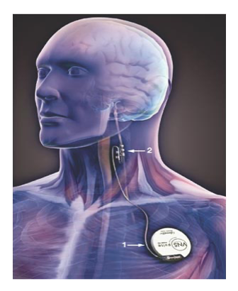
VNS is placed in patient. Generator is implanted in the upper chest; a lead with electrons is placed adjacent to the segment of the vagus nerve.
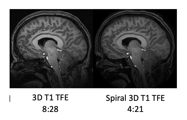
Example of a conventional 3D TFE compared to a spiral 3D TFE.
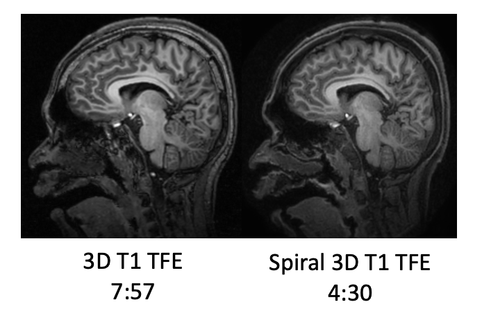
Example of a conventional 3D TFE compared to a spiral 3D TFE