Breathing Artifacts in Liver MRI: Emerging Solutions
1Department of Radiology, University of Yamanashi, Chuo, Japan
Synopsis
Many studies have attempted to reduce motion artifacts in the liver over the years. However, it is still challenging to develop robust and practical methods to address this problem because of the complicated nature of motion artifacts. Recently, the deep learning approach has been used to achieve excellent image processing results. This talk provides an overview of deep learning-based methods to address breathing artifacts in the liver.
Purpose
This talk aims to provide an overview of state-of-art methods to address breathing artifacts in the liver.Introduction
Abdominal MRI is very sensitive to motion, which is mainly caused by breath-hold failure during scans. For example, it is well known that the arterial phase of gadoxetate-enhanced MRI exhibits severe motion artifact1. Many approaches have been proposed to minimize motion artifacts. Respiratory triggering, which is a method of synchronous data acquisition based on breathing patterns provided by bellows or navigator echoes, is effective for minimizing motion artifacts2. This approach is widely used because of its straightforward mechanism. However, the limited data acquisition window of triggering leads to long scan times. Therefore, it is challenging to use the triggering approach for dynamic imaging that requires fast acquisition. Compressed sensing (CS) is a fast imaging approach utilizing underlying sparsity and random sampling, for abdominal MRI. It reduces the scan time significantly, thus resulting in less breath-hold time3. It is generally combined with parallel imaging to enhance the reduction factor. Radial acquisition is achieved by rotating the readout direction, and it is robust to motion compared with conventional Cartesian imaging. Recently, advanced techniques using radial imaging with golden angle trajectory and CS have demonstrated free-breathing scans for DCE-MRI for the liver4. Although it has excellent performance against the respiratory motion, its high computational cost is still being discussed. Furthermore, retrospective motion correction approaches are under intensive investigation5.Deep learning (DL), which extracts signal features using complicated non-linear processing, has received considerable attention for reducing motion artifacts6. It is a machine learning technique based on the neural network and is widely used for various purposes such as classification problems and image processing. Here, we introduce and describe emerging solutions, mainly focusing on DL to reduce motion artifacts.
DL-based motion artifact reduction
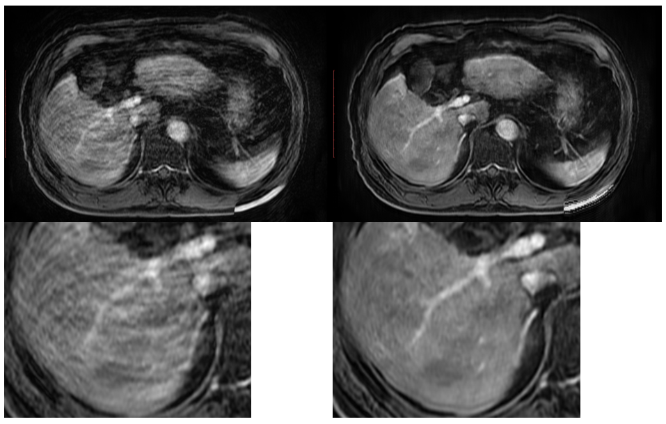
Many DL-based applications have been proposed to remove motion artifacts for the brain7-9, c-spine10, liver11, 12, upper abdomen13, and cardiac14 imaging. Jiang et al. proposed a motion artifact reduction technique using a GAN-based network with U-net as a generator network for abdominal MRI13. They demonstrated that the GAN approach successfully removes artifacts for gradient-echo and fast spin-echo sequences. Tamada et al. proposed a motion artifact reduction for DCE-MRI of the liver using a CNN-based network with multichannel input and output11. The datasets used for training and validation were generated by simulating respiratory motion. The simulation was achieved by adding random and periodical phase errors in the k-space data. The proposed network successfully reduced artifacts of arterial phase images.Limitations
Although some promising results have been obtained, there are challenges in removing artifacts using DL. It is challenging to prepare a pair of datasets of images with and without artifact, for training because of the misregistration among acquired images. Instead of using acquired images, simulated images can be used. However, it is technically challenging to simulate abdominal motion with high accuracy because of its complicated deformable motion. Further, artifacts induced by various factors such as cardiac and hardware imperfection can be observed in practical imaging in addition to motion artifacts. The generalization of the DL network is also concern. Because many different kinds of a sequence are used depending on vendors and facilities, a limited number of datasets can lead to the inappropriate training of the network.Acknowledgements
No acknowledgement found.References
1. Motosugi U, Bannas P, Bookwalter CA, Sano K, Reeder SB. An investigation of transient severe motion related to gadoxetic acid–enhanced MR imaging. Radiology 2016; 279:93-102.
2. Chavhan GB, Babyn PS, Vasanawala SSJR. Abdominal MR imaging in children: motion compensation, sequence optimization, and protocol organization 2013; 33:703-719.
3. Zhang T, Chowdhury S, Lustig M, et al. Clinical performance of contrast enhanced abdominal pediatric MRI with fast combined parallel imaging compressed sensing reconstruction 2014; 40:13-25.
4. Feng L, Grimm R, Block KT, et al. Golden‐angle radial sparse parallel MRI: combination of compressed sensing, parallel imaging, and golden‐angle radial sampling for fast and flexible dynamic volumetric MRI 2014; 72:707-717.
5. Cheng JY, Alley MT, Cunningham CH, Vasanawala SS, Pauly JM, Lustig MJMrim. Nonrigid motion correction in 3D using autofocusing withlocalized linear translations 2012; 68:1785-1797.
6. Tamada D. Noise and artifact reduction for MRI using deep learning. arXiv preprint arXiv:200212889 2020.
7. Pawar K, Chen Z, Shah NJ, Egan GF. Moconet: Motion correction in 3D MPRAGE images using a convolutional neural network approach. arXiv preprint arXiv:180710831 2018.
8. Duffy BA, Zhang W, Tang H, et al. Retrospective correction of motion artifact affected structural MRI images using deep learning of simulated motion 2018.
9. Johnson PM, Drangova M. Motion correction in MRI using deep learning. ISMRM Scientific Meeting & Exhibition, Honolulu, 2018; 4098.
10. Lee H, Ryu K, Nam Y, Lee J, Kim D-H. Reduction of respiratory motion artifact in c-spine imaging using deep learning: Is substitution of navigator possible? ISMRM Scientific Meeting & Exhibition, 2018; 2660.
11. Tamada D, Kromrey M-L, Ichikawa S, Onishi H, Motosugi U. Motion Artifact Reduction Using a Convolutional Neural Network for Dynamic Contrast Enhanced MR Imaging of the Liver. Magnetic Resonance in Medical Sciences 2020; 19:64-76.
12. Tamada D, Onishi H, Motosugi U. Motion Artifact Reduction in Abdominal MR Imaging using the U-NET Network. ICMRM and Scientific Meeting of KSMRM, Seoul, Korea, 2018; PP03–11.
13. Jiang W, Liu Z, Lee K-H, et al. Respiratory motion correction in abdominal MRI using a densely connected U-Net with GAN-guided training. arXiv preprint arXiv:190609745 2019.
14. Oksuz I, Clough J, Ruijsink B, et al. Detection and Correction of Cardiac MRI Motion Artefacts During Reconstruction from k-space. International Conference on Medical Image Computing and Computer-Assisted Intervention, 2019; 695-703.
Figures