How Can CNNs Improve the Quality of My Acquired MRS/MRSI Data?
1UT Southwestern, Dallas, TX, United States
Synopsis
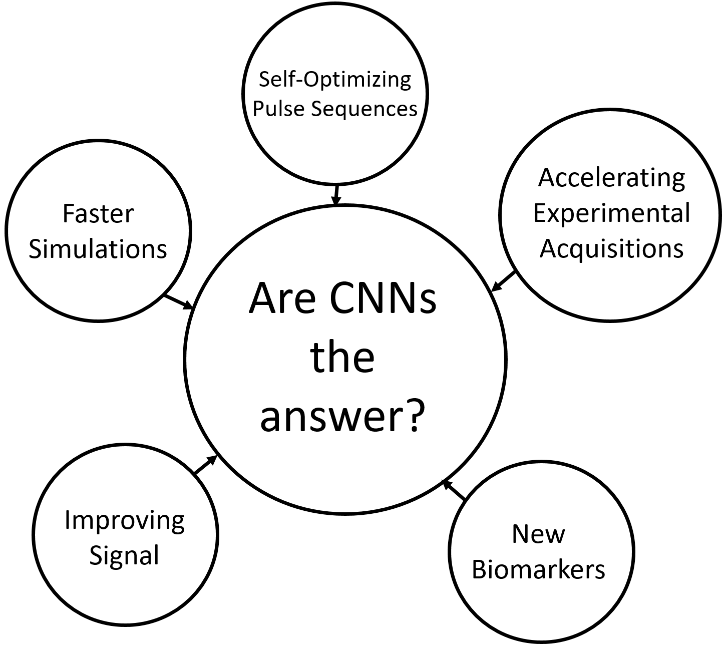
Deep learning is a new and exciting frontier for magnetic resonance spectroscopy (MRS) and spectroscopic imaging (MRSI) scientists. With this new technology, it may be possible to improve a many active areas of research, including simulations, pulse sequence design, experimental acquisitions, reconstruction, quantitation, and clinical outcome correlation. However, just like any new technology, deep learning has strengths and weaknesses that need to be understood before large-scale implementation. This presentation will explore how deep learning as improved the field of MRS and MRSI, and how it will continue to do so in the future.
Background
Magnetic resonance spectroscopy (MRS) and spectroscopic imaging (MRSI) are powerful tools capable of providing valuable metabolic information from a myriad of tissues in vivo[1,2]. Technological advancements over the past three decades have been very impressive and have led to improvements in pulse sequence design, experimental acquisition, reconstruction, and quantitation of results. For example, many multi-slice techniques with high spatial resolutions for MRSI demonstrate excellent agreement with anatomical magnetic resonance images (MRI) when comparing metabolite images and T1-weighted or T2-weighted images[3,4]. Also, several techniques exist that are now capable of detecting markers associated with genetic mutations[5,6]. Even with all of these developments, MRS and MRSI are still under-utilized in clinical workflows, mainly due to the longer acquisition times and technical difficulties of the methods. Recently, deep learning has become a popular technology that has had a large impact on medical imaging[7]. In particular, convolutional neural networks (CNNs)[8] have made great advancements in aiding diagnosis, treatment evaluation, and monitoring patient outcome. Supervised learning, where a model learns features from labelled data, has been the main focus in medical imaging, where CNNs are trained to perform of a variety of classification or transformation tasks based on previously collected data. In this presentation, we will discuss how CNNs have been implemented for MRS and MRSI, and will also delve into other potential applications of this technology that will most likely be explored in the future.Syllabus/Discussion
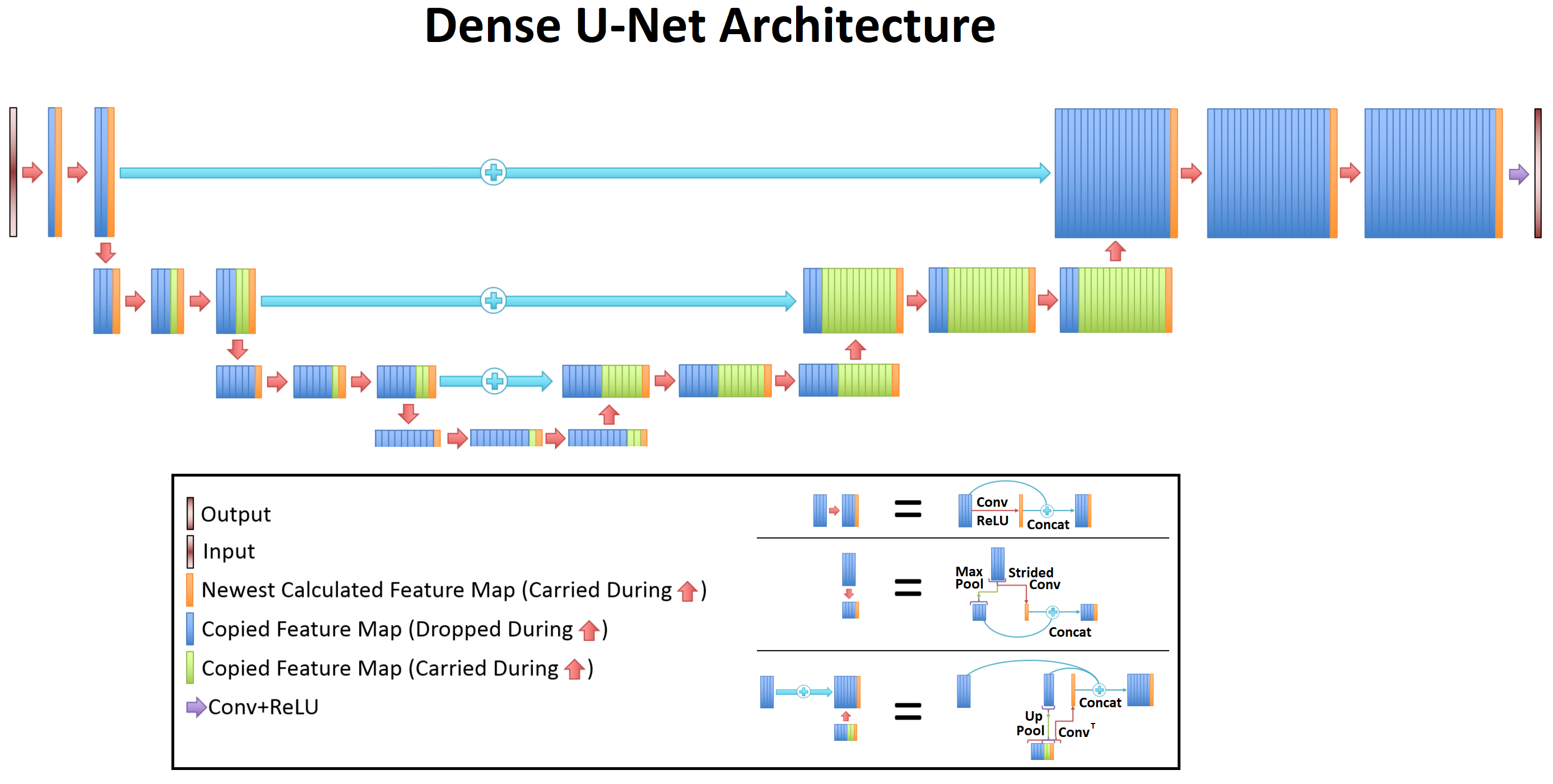
We will first briefly discuss MRS and MRSI, mainly the different aspects of the field where development is important: simulation, pulse sequence design, hardware, experimental acquisition, reconstruction, quantitation, and clinical evaluation. Next, we will quickly review the theory of CNNs, including the strengths and weaknesses of this method. It is important to note that CNNs are not the answer to every problem, and that they require a large supply of diverse data to be useful. Then, we will go through published studies that have used CNNs to improve the quality of acquired MRS and MRSI data. Finally, we will conclude by discussing future applications where CNNs may be helpful or find limited use.Acknowledgements
I would like to acknowledge all of the members of the MAIA laboratory at UT Southwestern, as well as all of my co-authors for their help and input.References
[1] Soares, D. P., and M. Law. "Magnetic resonance spectroscopy of the brain: review of metabolites and clinical applications." Clinical radiology 64, no. 1 (2009): 12-21.
[2] Costello, L. C., RB, and Franklin, and P. Narayan. "Citrate in the diagnosis of prostate cancer." The prostate 38, no. 3 (1999): 237-245.
[3] Hangel, G., Jain, S., Springer, E., Hečková, E., Strasser, B., Považan, M., Gruber, S., Widhalm, G., Kiesel, B., Furtner, J. and Preusser, M., 2019. High-resolution metabolic mapping of gliomas via patch-based super-resolution magnetic resonance spectroscopic imaging at 7T. Neuroimage, 191, pp.587-595.
[4] Posse, S., Otazo, R., Dager, S.R. and Alger, J., 2013. MR spectroscopic imaging: principles and recent advances. Journal of Magnetic Resonance Imaging, 37(6), pp.1301-1325.
[5] Choi, C., Ganji, S.K., DeBerardinis, R.J., Hatanpaa, K.J., Rakheja, D., Kovacs, Z., Yang, X.L., Mashimo, T., Raisanen, J.M., Marin-Valencia, I. and Pascual, J.M., 2012. 2-hydroxyglutarate detection by magnetic resonance spectroscopy in IDH-mutated patients with gliomas. Nature medicine, 18(4), p.624.
[6] Andronesi, O.C., Kim, G.S., Gerstner, E., Batchelor, T., Tzika, A.A., Fantin, V.R., Vander Heiden, M.G. and Sorensen, A.G., 2012. Detection of 2-hydroxyglutarate in IDH-mutated glioma patients by in vivo spectral-editing and 2D correlation magnetic resonance spectroscopy. Science translational medicine, 4(116), pp.116ra4-116ra4.
[7] LeCun, Y., Bengio, Y. and Hinton, G., 2015. Deep learning. nature, 521(7553), pp.436-444.
[8] Krizhevsky, A., Sutskever, I. and Hinton, G.E., 2012. Imagenet classification with deep convolutional neural networks. In Advances in neural information processing systems (pp. 1097-1105).
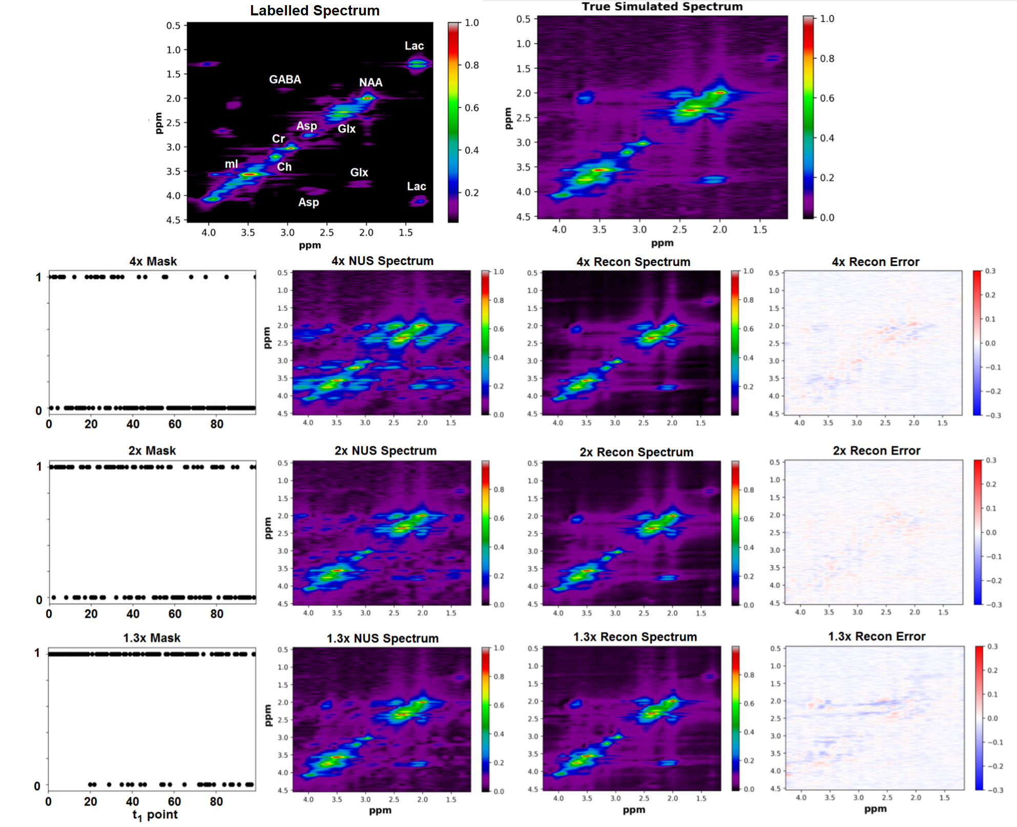
[9] Iqbal, Z., Nguyen, D., Hangel, G., Motyka, S., Bogner, W. and Jiang, S., 2019. Super-Resolution 1H Magnetic Resonance Spectroscopic Imaging utilizing Deep Learning. Frontiers in oncology, 9.
[10] Iqbal, Z., Nguyen, D., Thomas, M.A. and Jiang, S., 2018. Acceleration and quantitation of localized correlated spectroscopy using deep learning: a pilot simulation study. arXiv preprint arXiv:1806.11068.
Figures