4873
Diagnostic efficiency of DBT compared to conventional MRI and FFDM in diagnosing breast cancer
Fan WenWen1, Fan WenWen1, Ou Yang Han2, Zhao XinMing2, Zhang HongMei2, and Xie LiZhi3
1Department of Imaging Diagnosis, National Cancer Center/Cancer Hospital, Chinese Academy of Medical Science and Peking Union Medical College, BeiJing, China, 2National Cancer Center/Cancer Hospital, Chinese Academy of Medical Science and Peking Union Medical College, BeiJing, China, 3MR Research, GE Healthcare, BeiJing, China
1Department of Imaging Diagnosis, National Cancer Center/Cancer Hospital, Chinese Academy of Medical Science and Peking Union Medical College, BeiJing, China, 2National Cancer Center/Cancer Hospital, Chinese Academy of Medical Science and Peking Union Medical College, BeiJing, China, 3MR Research, GE Healthcare, BeiJing, China
Synopsis
The aim of this study was to evaluate the technical characteristics and diagnostic efficiency of Digital Breast Tomosynthesis (DBT) compared to conventional MRI approach and full-field digital mammography (FFDM) in screening breast cancer (BC) patients. Briefly, 253 female patients who underwent Digital Mammary Gland 3D Tomosynthesis, MR scan and FFDM were enrolled in this study. Briefly, the DBT, FFDM and MRI showed a sensitivity of 90.9%, 85.6% and 94.1%; specificity of 89.7%, 80.7% and 83.3%; and coincidence rate of 89.3%, 83.4% and 89.7%, respectively, and the difference was statistically significant (p<0.001).
Introduction
Over the recent years, the incidence rate of breast cancer (BC) in China has been rapidly increasing. Early diagnosis of BC remains a critical step in selecting the best treatment option and prolonging the patient’s survival. So far, different tests have been developed for the BC diagnosis. Conventional methods include digital mammography, followed by Breast Ultrasound and breast MRI. DBT is new type of pseudo-3D imaging method used for screening breast, which results in better discrimination of tissue structures and potentially improved visualization of cancer. Recent data have suggested that DBT can improve both specificity and sensitivity of imaging in BC screening, leading to higher rates of detected cancers with fewer false-positives (which can be easily observed by digital mammography).Methods
A total of 253 female patients were randomly selected and enrolled in this study. All patients underwent Digital Mammary Gland 3D Tomosynthesis and MR scan; pathological and imaging data were compared. Digital mammography was performed under standard mode of Hologic mammary gland machine to obtain the corresponding 2D and 3D images. 3.0T MR system (MR 3.0T Twinspeed signa hdxt, GE Healthcare, America) was preformed using multiple-b-value diffusion-weighted imaging (DWI) scans; the following imaging parameters were used for FS AX T2 IDEAL sequence (FOV=33×33cm, slice thickness=5.0mm, Echo Train Length=19, bandwidth=62.5, matrix=320×224, TE=85ms, TR=5440ms). The following imaging parameters were used for STIR AX DWI sequence (FOV=40×40cm, slice thickness=8.0mm, TE=Minimum, TR=4000, Inv. Time=160, NEX=2.0, matrix=128×128). The following imaging parameters were used for AX VIBRANT+C sequence (FOV=30×30cm, slice thickness=2.0mm, flip angle=15, bandwidth=62.5, matrix=388×256).GE AW4.4 workstation was used to calculate the value of ADC, ADC standard, D, D*, f, and to generate time-signal intensity curve (TIC). All parameters between benign and malignant breast lesions were compared by Mann-Whitney U test. Finally, we performed an analysis of breast cancer focus based on 114 cases of female patients identified by histopathology. Receiver operating characteristic curve analysis evaluated the diagnostic performance of different parameters.Results & Discussion
A total of 130 patients were diagnosed with BC using DBT. Among the patients, 96 had negative results after pathological examination of the tissue. These data suggested sensitivity rate of 90.9%, specificity of 89.7%, and accuracy of 89.3% of DBT in detecting BC (Table 1; Figure 1 and 2). Moreover, 119 and 142 cases were diagnosed with BC using FFDM and MRI, respectively; where 92 and 85 cases had negative results, which suggested that FFDM and MRI have a sensitivity of 85.6% and 94.1%, specificity of 80.7% and 83.3%, and the coincidence rate of 83.4% and 89.7%, respectively . More importantly, the difference between the groups was statistically significant (p<0.001), which was consistent with previous studies [1,2].Conclusion
These data suggested that DBT offers better specificity, but lower sensibility and accuracy in detecting breast cancer compared to the MRI approach. However, compared to MRI, it implies lower cost and fewer contraindications. In addition, both MRI and DBT are considered as superior choices compared to conventional FFDM method. Yet, compared to MRI, they lead to lower cost and fewer contraindications.Acknowledgements
No acknowledgement found.References
[1] Park A M, Franken EA, Garg M, et al. Breast tomosynthesis: Present considerations and Future applications [J],Riadiographics, 2007(27):231-240.[2] Fischmann A, Siegmann KC, Wersebe A, et al. Comparison of full-filed digital mammography and full-screen mammography :image quality and lesion detection [J]. Radiology ,2005,78(928):312-317Figures
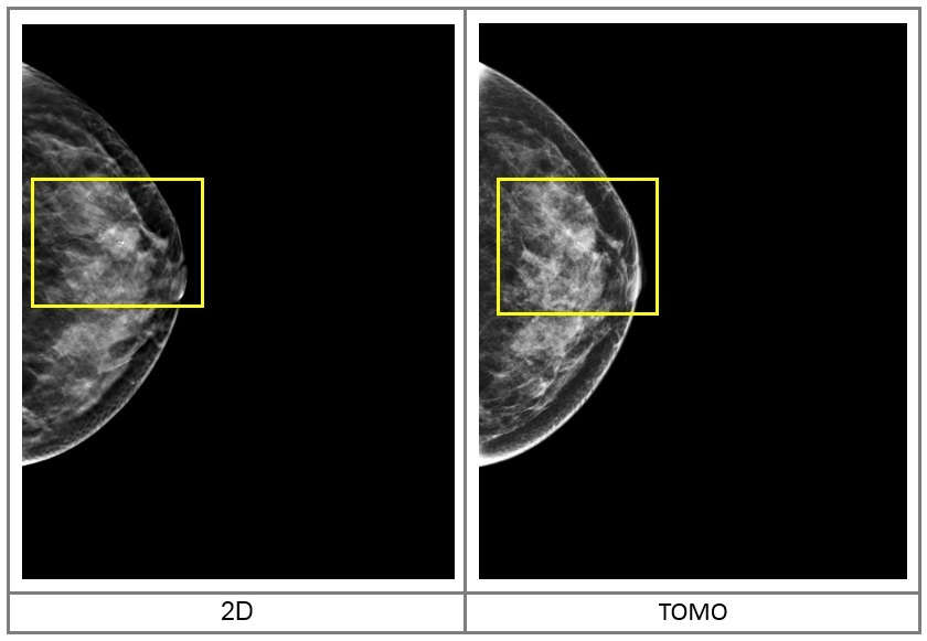
A single slice of the 3D tomosynthesis acquisition showing spiculated margins of the mass.
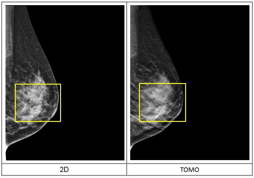
2D image and 3D Tomosynthesis Slices
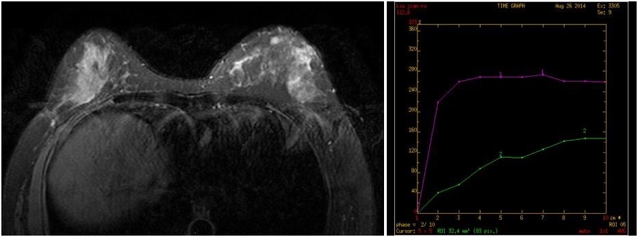
The left breast quadrant showing a lobulated nodule, Edge visible burr, 1.1cmx0.8cmx1.6cm in size, T2WI/FS is signal, DWI diffusion limited, Enhanced scanning showed obvious enhancement, Time signal curve is platform type.
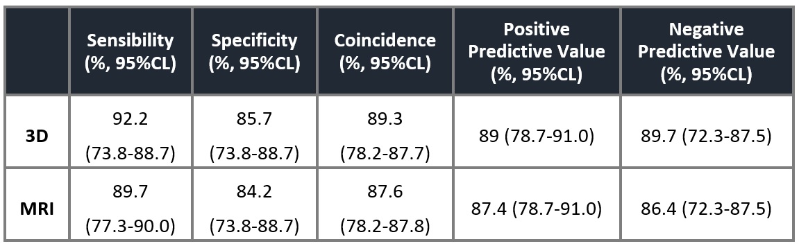
Sensibility, specific and accuracy of detecting BC with 2D, 3D and MRI