4872
A Pilot Evaluation of Intravoxel Incoherent Motion Imaging Features of Stage T1 HER2-positive breast carcinomas1The First Affiliated Hospital of Dalian Medical University, Dalian, China, 2GE Healthcare, MR Research China, Beijing, China
Synopsis
Intravoxel incoherent motion (IVIM) imaging provides quantitative measurement of ADCslow for cellularity and ADCfast and ffast for vascularity. The parameters derived from IVIM can distinguish lesions of different aggressive pathological subtypes of Stage T1 breast carcinomas[1]. This study concerned clinical features, perfusion as well as diffusion parameters using IVIM imaging and then compared these parameters from conventional MR images on the classification of Stage T1 HER2-positive and HER2-negative breast carcinoma lesions .
Purpose
To investigate the use of IVIM parameters in predicting Stage T1 HER2-positive breast carcinoma patients.Introduction
For stage T1 HER2-positive breast cancer patients, some clinical features were present as factors for the prognosis inculding axillary lymph node metastasis etc[1]. There were still challenges of discrination using current methods including conventional MRI and DWI.Compared to conventional diffusion weighted imaging (DWI), IVIM has the ability to separate diffusion from perfusion and hence has the potential to better assess the tissue celluarity[2]. In this work the use of IVIM in predicting predicting Stage T1 HER2-positive breast carcinoma is investigated.Methods
In this prospective study, a total of 51 female patients with stage T1 breast carcinomas in pathology were enrolled. Ethical approval and informed consent forms were obtained. The patients were divided into two groups: group 1 included 29 patients with HER2-positve carcinomas and group 2 included 22patients with HER2-negative carcinomas. All the participants underwent MR exams including IVIM, T1-weighted, T2-weighted and contrast-enhanced MRI. Clinical features (age, axillary lymph node metastasis), tumor size, IVIM parameters (ADCslow, ADCfast and ffast) and MRI features of lesions including Fibrograndular tissue (FGT), background parenchymal enhancement (BPE), morphological manifestations were obtained. The parameters were compared between two groups and the diagnostic performance was quantified with ROC curve. A multivariate logistic regression model was developed to identify features that were independently predictive for tumor HER2 expression.Results
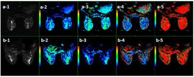
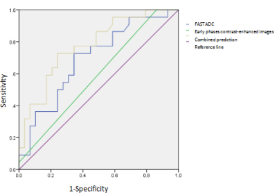
Images of a HER2-positive and a negative patient are shown in Figure 1. There was no significant difference in age, axillary lymph node metastasis, FGT, BPE, ADCslow and f between two groups (p=0.054, 0.062, 0.153, 0.226, 0.924, 0.052). Stage T1 Her2-positive group can be significantly differentiated from the lesions of HER2-negative group based on shapes of early phases contrast-enhanced images and ADCfast (p=0.037, 0.019). The highest area under the curve (AUC) was acquired by combining shapes on early phases contrast-enhanced inages and ADCfast with optimal threshold of 0.777(Figure 2).Discussion and Conclusion
HER2 expression is an important factor impacting the prognosis of early breast cancer[3]. For Stage T1 HER2-positive breast carcinomas patients, the presence of axillary lymph nodes metastasis is an independent factor for the prognosis[4]. Biomarkers that may predict of the prognosis of patients diagnosed with T1 HER2-positive breast carcinomas hold great clinical value. This study suggests that the parameters derived from IVIM may distinguish lesions of Stage T1 HER2-positive and HER2-negative breast carcinoma lesions, taking into account of stage of menstrual cycle and status of axillary lymph nodes. These findings support the pre-operative MRI for the discrimination of HER2 expression of Stage T1 breast carcinomas.Acknowledgements
This work was supported by the grant of the Basic Scientific Research Projects of the Universities in Liaoning Province (5061104) and The Provincical Natural Science Foundation of China (2019-ZD-0907) .References
[1] Lina Z, Jianyun K, Qingwei S, et al. Intravoxel incoherent motion imaging in the prediction of aggressiveness of T1 breast carcinoma patients. ISMRM,2019, May13.
[2] Yuan Y ,Hu SN,Gao J, Expression discordances and clinical values of ER, PR, HER-2 and Ki-67 in primary and metastatic breast cancer. Zhonghua Zhong Liu Za Zhi.2019 ,41(9) :681-685.
[3] Ovcaricek T, Takac I,Matos E,et al.Multigene expression signatures in early hormone receptor positive HER 2 negative breast cancer. Radiol Oncol. 2019, 24 ,53(3) :285-292.
[4] Zhang Y ,Li J ,Fan Y,et al.Risk factors for axillary lymph node metastases in clinical stage T1-2N0M0 breast cancer patients. Medicine (Baltimore).2019,98(40) :e17481.
Figures

