4859
Diffusion Kurtosis Imaging and Intravoxel Incoherent Motion MR Imaging in the Differentiation of Sinonasal Malignant Tumors
Zuohua Tang1, Zebin Xiao1, Peng Wang1, and Zhongshuai Zhang2
1Radiology, Fudan University Eye & ENT Hospital, Shanghai, China, 2MR Scientific Marketing, Siemens Healthcare, Shanghai, China
1Radiology, Fudan University Eye & ENT Hospital, Shanghai, China, 2MR Scientific Marketing, Siemens Healthcare, Shanghai, China
Synopsis
A reliable differentiation of different histological types of sinonasal malignancies is usually difficult by using conventional CT and MR imaging but plays an important role in determining the treatment strategies for the patients. This is the first study which combined DKI and IVIM to distinguish among four histological types of sinonasal malignant tumors, including squamous cell carcinomas (SCCs), olfactory neuroblastomas (ONBs), malignant melanomas (MMs), and lymphomas. Our study showed that these sinonasal malignant tumors had distinct profiles in DKI and IVIM parameters, determined by different proportions of tissue complexity, capillary perfusion, or water diffusion components within tumor.
Synopsis
A reliable differentiation of different histological types of sinonasal malignancies is usually difficult by using conventional CT and MR imaging but plays an important role in determining the treatment strategies for the patients. This is the first study which combined DKI and IVIM to distinguish among four histological types of sinonasal malignant tumors, including squamous cell carcinomas (SCCs), olfactory neuroblastomas (ONBs), malignant melanomas (MMs), and lymphomas. Our study showed that these sinonasal malignant tumors had distinct profiles in DKI and IVIM parameters, determined by different proportions of tissue complexity, capillary perfusion, or water diffusion components within tumor.Background and purpose
Histopathologically, malignant tumors in the nasal cavity usually include SCCs, ONBs, MMs, and lymphomas. In many cases, these four malignant tumors cannot be easily differentiated on CT and conventional MRI. Diffusion kurtosis imaging (DKI) has been proven to be capable of characterizing the tumoral microstructure and complexity which are highly varied in sinonasal malignancies, and therefore holds great potential to indicate some histological types of malignant tumors in the sinonasal region1, 2. Intravoxel incoherent motion (IVIM) has emerged as a useful method to non-invasively assess the neovascularization of malignant tumors that is a critical pathological factor for tumor growth, invasion, and metastasis. Thus, estimation of hemodynamic differences using IVIM may also be predictive of sinonasal malignancies in some histological types.1, 2 Hence, the combination of DKI and IVIM can comprehensively evaluate these three distinctive physiological and pathological features, including tumor cellularity, heterogeneity and perfusion, and thus may be helpful for the differential diagnosis of different types of sinonasal malignancies. Therefore, our study aimed to evaluate whether the combined use of diffusion parameters derived from DKI and IVIM models was applicable for differentiating the four histological types of sinonasal malignant tumors, including SCCs, ONBs, MMs and lymphomas.Methods
65 patients with sinonasal malignancies who underwent DKI and IVIM on a 3T MR scanner (MAGNETOM Verio, Siemens Healthcare, Erlangen, Germany) were enrolled in this retrospective study, including 27 squamous cell carcinomas (SCCs), 13 olfactory neuroblastomas (ONBs), 14 malignant melanomas (MMs). and 11 lymphomas. The detailed parameters were as follows: TR/TE = 5200/83 msec, δ = 27.4 msec, Δ = 39.4 msec, number of averages = 2, acquisition matrix = 120 × 120; field of view (FOV) = 220 mm, slice thickness = 5 mm, intersection gap = 5 mm, parallel imaging acceleration factor = 2; 14 different b values ranging from 0 to 2500 sec/mm2 were used (b = 0, 50, 100, 150, 200, 250, 300, 350, 400, 800, 1000, 1500, 2000, and 2500 sec/mm2). The IVIM and DKI processing were performed using custom-written scripts in MATLAB (version R2016a; MathWorks, Natick, Mass) to provide ADC, D, D*, f, Dk and K parametric maps on a pixel-by-pixel basis.3 Kruskal-Wallis and Dunn multiple comparison tests with Bonferroni correction, receiver operating characteristic curve, and logistic regression analyses were used for statistical analysis.Results
Lymphomas demonstrated highest K values but lowest Dk, D, D*, f and f∙D* values among these four malignant tumors. ONBs exhibited high K values and MMs had highest D*, f and f∙D* values. The cutoff value ≤ 0.887 × 10⁻³ mm2/s for f∙D* provided a sensitivity, specificity and an accuracy of 100%, 98.1% and 98.5%, respectively, for differentiating lymphomas from other three entities. The combination of f∙D* and D values showed a sensitivity of 92.9% and a specificity of 92.5% for the discrimination of MMs from ONBs and SCCs. The K value was useful for differentiating ONBs from SCCs, with a threshold value of 0.942 (sensitivity, 84.6%; specificity, 63.0%). All the results are summarized in Figure 1.Conclusion
The combined use of DKI and IVIM is helpful for differentiating among four histological types of sinonasal malignant tumors.Acknowledgements
No acknowledgement found.References
1. Simons SA, Bridge JA, Leon ME. Sinonasal small round blue cell tumors: An approach to diagnosis. Seminars in diagnostic pathology 2016;33:91-103 2. Thompson LD. Small round blue cell tumors of the sinonasal tract: a differential diagnosis approach. Modern pathology : an official journal of the United States and Canadian Academy of Pathology, Inc 2017;30:S1-s26 3. Xiao Z, Zhong Y, Tang Z, et al. Standard diffusion-weighted, diffusion kurtosis and intravoxel incoherent motion MR imaging of sinonasal malignancies: correlations with Ki-67 proliferation status. European radiology 2018;28:2923-2933Figures
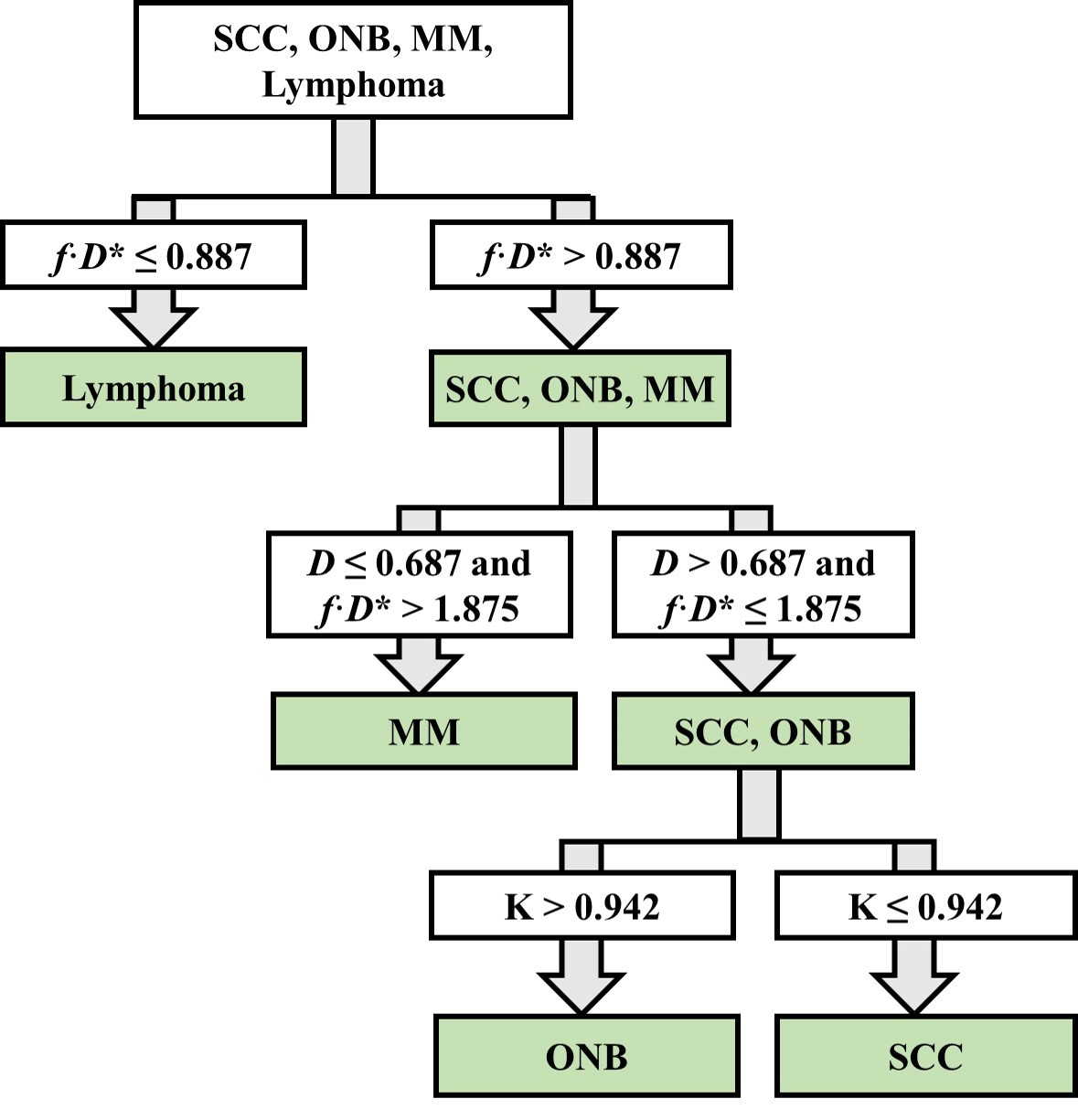
Fig. 1: Stepwise differentiation of four sinonasal malignant tumours, including lymphomas, olfactory neuroblastomas, squamous cell carcinomas and malignant melanomas using DKI and IVIM parameters.