4816
The correlation analysis of 3D APT with the parameters of 3D MR spectroscopy in glioma patients: an image-guided intraoperative pilot study
Xiaoluo Zhang1, Dongxiao Zhuang1, Jilei Zhang2, Jianqing Sun2, Weibo Chen2, and Jinsong Wu1
1Glioma Surgery Division, Neurologic Surgery Department, Huashan Hospital, Fudan University, Shanghai, China, 2Clinical Science, Philips Healthcare, Shanghai, China
1Glioma Surgery Division, Neurologic Surgery Department, Huashan Hospital, Fudan University, Shanghai, China, 2Clinical Science, Philips Healthcare, Shanghai, China
Synopsis
Our study is the first research to investigate the relationship between 3D APT and 3D MRS on individual level. The APTw values were correlated with the metabolite concentrations or ratios of MRS, especially Cho concentration. We proposed that 3D APT can be potentially used in intra-operation system to guide surgical biopsy and tumor resection.
Introduction
Previous study demonstrated that MR spectroscopy may be a helpful tool for tumor resection during glioma surgery and can be used to guide surgical biopsy for intraoperative neuronavigational systems[1]. 3D Amide proton transfer (APT) imaging, as a novel chemical exchange saturation transfer (CEST)‐based molecular MRI technique, has been recently proposed as an important biomarker for brain glioma grading, and reflect the cellular proliferation levels that correlate with Ki-67[2]. In current study, we investigate the relationship among 3D APT and the parameters of 3D MR spectroscopy on individual level, and propose that 3D APT can be potentially used in intraoperation system to guide surgical biopsy and tumor resection.Methods
Institutional Review Board approval and written informed consent were obtained. Conventional MRI images, 3D APT, 3D MRS data and 3D T1-weighted data were conducted using an MR-OR system (Ingenia 3.0T, Philips Healthcare, Best, The Netherland). Four glioma patients were recruited in current study with 2 astrocytomapatients (WHO II and WHO III) and 2 glioblastoma patients (WHO IV). The pipeline of data process was implemented in Figure 1. The co-registration was performed between quantitative APT weighted images (APTw) and 3D MRS based on 3D T1-weighted image. The APTw images were resampled to the space of MRS data. The slice which includes maximum tumor area within 3D MRS data were selected to perform correlation analysis. The corresponding APTw values within the region of interest, which were drawn on solid portion of tumor, were extracted for each voxels and then correlated with the choline (Cho), creatine (Cr) and N-acetylaspartate (NAA) concentration, as well as the Cho/Cr and Cho/NAA ratios using voxel-wise methods.Results
The results of correlation analysis between APTw values within solid tumor tissue and the parameters of 3D MRS in patients with glioma were exhibited in Table1 and Figure 2. The APTw values of 2 patients with astrocytoma and 1 patients withglioblastomaare significantly positive correlation with Cho concentration (p<0.05). It is noteworthy that the APTw values of one patient with astrocytoma are significantly positive correlation with Cho/NAA ratio, and negative correlation with NAA concentration (p<0.05). However, the APTw values of one glioblastoma patient are slightly positive correlation with Cho concentration (p = 0.186) and Cho/NAA ratio (p=0.262), and negative correlation with NAA concentration (p=0.87). Figure 3 exhibited that the fusion of 3D APT and 3D T1w images can be integrated into the navigation system and get excellent results.Discussion
Our study is the first research to investigate the relationship between 3D APT and 3D MRS on individual level. The APTw values were correlated with the metabolite concentrations or ratios of MRS, especially Cho concentration which considered to be bio-marker for accelerated cell proliferation and reflect increased protein expression and proteolysis in tumor tissue. Our results suggested that the APT with higher resolution and coverage size for tumor tissue than 3D MRS can be served as a potential tool to guide surgical biopsy and tumor resection in intraoperation system.Acknowledgements
No acknowledgement foundReferences
1. Sasayama T, Kohta M, Tanaka K, et al. Surg-12. Intraoperative Magnetic Resonance Spectroscopy (imrs) For Glioma Surgery[J]. Neuro-Oncology, 2017, 19(Suppl 6): vi238.
2. Togao O, Hiwatashi A, Keupp J, et al. Amide proton transfer imaging of diffuse gliomas: effect of saturation pulse length in parallel transmission-based technique[J]. PloS one, 2016, 11(5): e0155925.
Figures
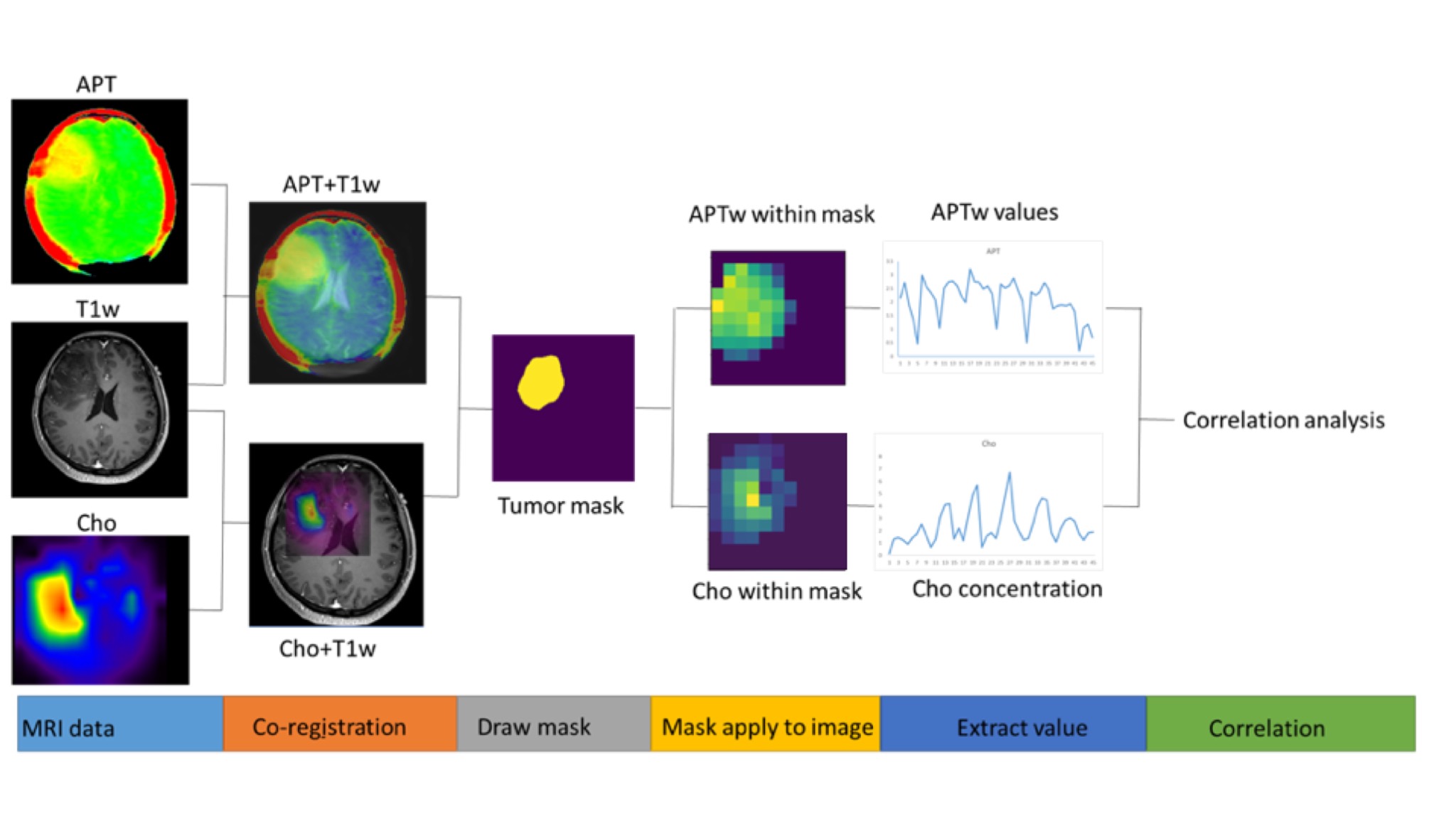
Figure 1. The pipeline of correlation analysis for APT and MRS.
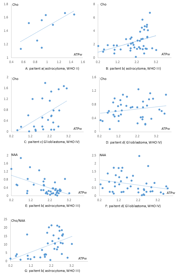
Figure 2. The scatter plot of correlation analysis between APTw values within solid tumor tissue and the metabolite concentrations or ratios of 3D MR spectroscopy in patients with glioma.
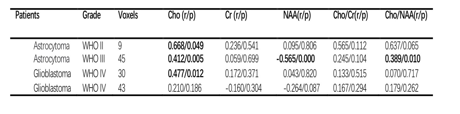
Table 1. The results of correlation analysis between APTw values within solid tumor tissue and the metabolite concentrations or ratios of 3D MR spectroscopy in patients with glioma.
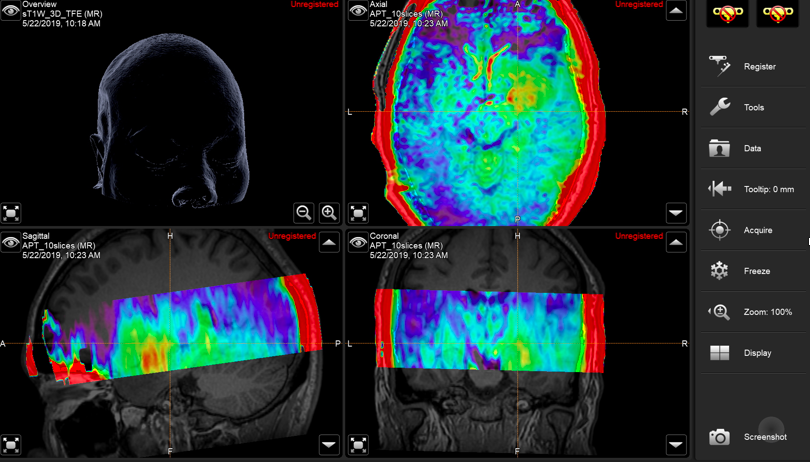
Figure 3.The fusion of 3D APT and 3D T1w images can be integrated into the navigation system to guide surgical biopsy and tumor resection