4803
Reproducibility of intravoxel incoherent motion MRI of pancreas tumor and healthy pancreas1CIBM-AIT, École polytechnique fédérale de Lausanne, Lausanne, Switzerland, 2Department of Radiology, Changhai Hospital of Shanghai, Shanghai, China
Synopsis
Intravoxel Incoherent Motion (IVIM) model has shown the potential of contrast free perfusion estimation in pancreatic diseases study, while IVIM quantification is challenging for conventional free-breathing DWI data due to the presence of high level noise. In this study, we demonstrated that motion correction and denoising can improve the sampling accuracy which allows for pixel-wise parametric maps with acceptable reproducibility.
Introduction
Diffusion weighted imaging (DWI) is a promising tool to facilitate pancreatic tumor detection and characterization (1). Among the diffusion reconstruction methods, the Intravoxel Incoherent Motion (IVIM) model becomes popular since it shows potential in providing perfusion related parameters without administration of contrast agents (2). Accurate estimation of perfusion parameters in IVIM model requires high SNR data, however, DWI in pancreas suffers from sampling error due to respiration and bowel peristalsis motion, the latter is unpredictable. In this study, we aim to investigate the possibility of tissue characterization in pancreatic tumor patients using free-breathing IVIM diffusion data after retrospective motion correction and noise removal. DWI metrics reproducibility was assessed in repeated scans. Considering the reproducibility analysis might be affected by the inherent heterogeneity of pancreatic adenocarcinoma, pancreas from healthy subjects were also included in the study.Methods
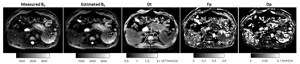
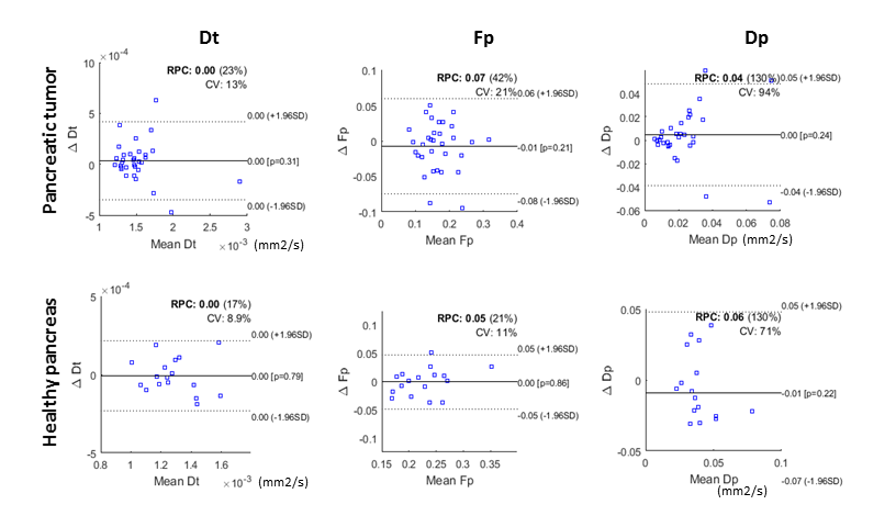
A total of 32 patients with pancreatic tumors and 17 healthy volunteers were scanned on a 3.0 T clinic system (GE Signa MR 750). Free-breathing 2D diffusion-weighted imaging (3-trace) was performed using axial single-shot EPI sequence with the following parameters: b = 0, 25, 50, 75, 100, 150, 200, 400, 600, 800 s/mm2, TE/TR = 76/4000 ms, 256×256 matrix (Interpolated), in plane 1.5×1.5 mm2, 24 slices with thickness of 6 mm and 1 mm gap. All subjects were scanned twice in the same session. After the first scan, each subject was requested to re-enter the scanning room and re-positioned for a repeated MRI scan. For data pre-processing, first, motion correction was achieved in the Advanced Normalization Tools (ANTs). In which, pair-wise deformable registration (3) with mutual information metric was performed for each diffusion-weighted volume, using b = 200 image as template. Motion corrected data was further filtered using local PCA based denoising (4) approach with a local patch radius of 2. The IVIM parameters were estimated from a segmented fitting approach: pure diffusion coefficient Dt was calculated from b ≥ 200 data using mono-exponential model; then estimated b = 0 map, perfusion fraction Fp and the pseudo-diffusion coefficient Dp were fitted using nonlinear least-square solver in Matlab. Region of interest (ROIs) were determined by user for each scan, either on healthy pancreas or within the pancreatic tumor, mean IVIM parameters in ROIs were used for reproducibility analysis. The 95% Bland-Altman limits of agreements and inter-subject coefficient of variation (CV) were reported.Results
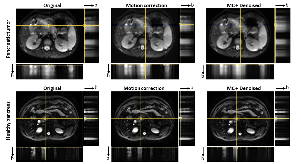
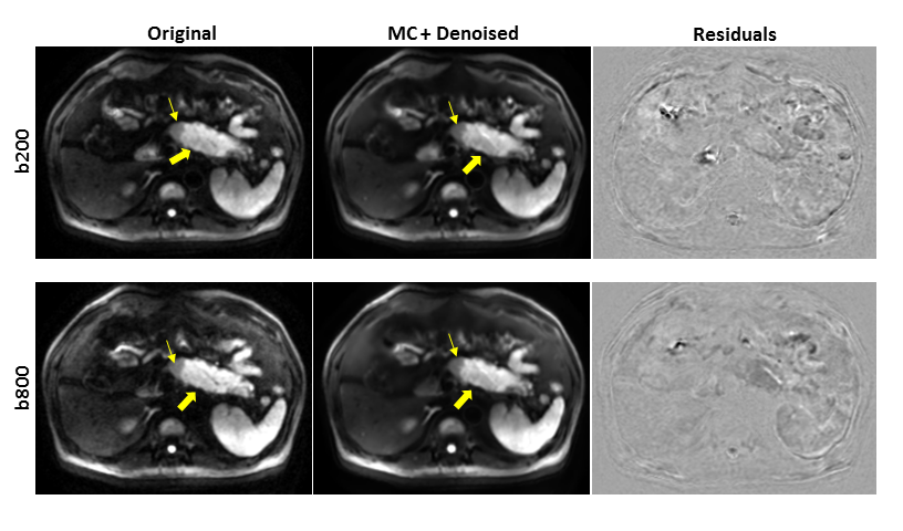
Non-linear deformable registration combing with local PCA based denoising have shown the potential of “freezing” the pancreas and pancreatic adenocarcinoma from both in plane and through plane motion artifact induced by peristalsis movement (Figure 1). The pancreas was less affected by respiration motion artifact at this anatomic level compared to the liver. Tissue boundary was well preserved after pre-processing, while contrast was slightly smoothed within tissue (Figure 2). Pixel-wise IVIM parameter maps were generated from two step fitting (Figure 3). In general, pancreatic adenocarcinoma has a lower perfusion fraction compare to adjacent pancreas tissue. Due to the limited sampling of b<100 data, the estimation of Dp is still challenging (CV>90% for tumor). Reproducibility of repeated scans was presented in Bland-Altman plot (Figure 4). Inter-subject CV for pancreatic tumor is generally higher than in healthy pancreas. In healthy pancreas, inter-subject CV is 8.9% for Dt and 11% for Fp.Discussion and conclusion
Though IVIM modeling has the potential ability to provide contrast free perfusion metrics in pancreatic cancer study, IVIM quantification is challenging due to the high sensitivity to noise and limited sampling in low b values. Previously a few studies have applied Bayesian methods (5,6) which have proven to be robust to noise, however the fitting procedure is time consuming. Here in this study, we demonstrated that the sampling error and low signal-to-noise ratio in conventional free-breathing DWI data can be partially recovered by motion correction and denoising steps.Acknowledgements
This work was supported by the Centre d’Imagerie BioMédicale (CIBM) of the UNIL, UNIGE, HUG, CHUV, EPFL and the Leenaardsand Louis-Jeantet Foundations, and the National Natural Science Foundation of China (81601468).References
1. Barral M, Taouli B, Guiu B, et al. Diffusion-weighted MR imaging of the pancreas: current status and recommendations. Radiology 2015;274:45–63.
2. Le Bihan D. What can we see with IVIM MRI? Neuroimage 2019;187:56–67.
3. Avants BB, Epstein CL, Grossman M, Gee JC. Symmetric diffeomorphic image registration with cross-correlation: Evaluating automated labeling of elderly and neurodegenerative brain. Med. Image Anal. 2008;12:26–41.
4. Manjon J, Coupe P, Concha L, et al. Diffusion Weighted Image Denoising Using Overcomplete Local PCA. PLoS One 2013;8:e73021.
5. Jalnefjord O, Andersson M, Montelius M, et al. Comparison of methods for estimation of the intravoxel incoherent motion (IVIM) diffusion coefficient (D) and perfusion fraction (f). Magn. Reson. Mater. Physics, Biol. Med. 2018;31:715–723.
6. Zhang L, Vishnevskiy V, Jakab A, Goksel O. Implicit modeling with uncertainty estimation for intravoxel incoherent motion imaging. Proc. - Int. Symp. Biomed. Imaging 2019;2019–April:1003–1007.
Figures