4762
3D amide proton transfer MR imaging for evaluation of hepatocellular carcinoma1The First Affiliated Hospital of Dalian Medical University, Dalian, China, 2Philips Healthcare, Beijing, China
Synopsis
Amide proton transfer weighted (APTw) imaging has been proved of great value in diagnoses of brain tumors, and has attracted increased interest for evaluation of abdomen diseases. Different regions of the hepatocellular carcinoma (HCC) are associated with different pathophysiological changes. The APTw-MRI was employed in this study to evaluate the protein and polypeptide molecular levels in different regions of HCC, and a significant difference was observed between APT values measured in the center of HCC and outside the tumor.
Purpose
To evaluate the protein and polypeptide molecular levels in different regions of hepatocellular carcinoma by APTw-MRI.Introduction
Amide proton transfer weighted (APTw) imaging is a recently developed contrast-agent-free MR technique that has been proved of great value in diagnoses of brain tumors[1,2]. And application of APTw-MRI for evaluation of abdomen diseases has attracted increased interest[3,4]. Different regions of the hepatocellular carcinoma (HCC) can be associated with different pathophysiological changes. In this study, the APTw-MRI was employed to evaluate the protein and polypeptide molecular levels in different regions of HCC.Methods
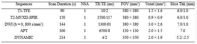
21 patients (15 males, 6 females) with hepatocellular carcinoma (clinical symptom and image preliminary diagnosis) were recruited (during May to October 2019), and underwent MR scans on a 3.0 T scanner (Ingenia CX, Philips Healthcare, Best, The Netherlands). MR sequences including T1-TFE, T2-MVXD-SPIR, DWI (b=800), APT, and DYNAMIC were used with parameters listed in Table 1. Patient exclusion criteria included: 1. missing from operation or puncture pathology, 2. the APTw-MR scan failed. Data from 15 patients were used after exclusion (12 males and 3 females). Fusion of T2w- and APTw-images was carried out by using the vendor-provided workstation (IntelliSpace Portal, Philips healthcare). Three circular regions of interest (ROI) were placed respectively at the center of hepatocellular carcinoma (avoid the lesion area of the capsule), around the hepatocellular carcinoma, and outside the tumor by two radiologists (Fig. 1). ROI sizes were 100, 120, and 120 mm2, respectively. The mean APT value of each region was calculated for all patients. The Mann-Whitney U test was used to detect difference of among APT values measured from different regions of hepatocellular carcinoma on platform of SPSS22.0 (IBM).Results
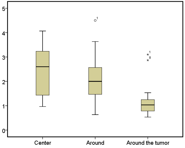
The mean APT values measured in the center of hepatocellular carcinoma, around and around the tumor were (2.40 ± 1.08)%, (2.08 ± 1.06)%, (1.23 ± 0.76)%, respectively. A significant difference was observed between APT values in the center of HCC and around the tumor (P = 0.005) (Fig. 2).Conclusion
In summary, the APTw-MRI show different APT values as well as different protein and polypeptide molecular levels in different regions of HCC. From the center of HCC to around the tumor, the APT value showed gradient changing. The APT values measured from the center HCC were significantly higher than that of around the tumor.Acknowledgements
No acknowledgements found.References
[1] Li B, Sun H, Zhang S, et al. Amide proton transfer imaging to evaluate the grading of squamous cell carcinoma of the cervix: A comparative study using 18F FDG PET. J Magn Reson Imaging. 2019;50(1):261-268.
[2] Takayama Y, Nishie A, Sugimoto M, et al. Amide proton transfer (APT) magnetic resonance imaging of prostate cancer: Comparison with Gleason scores. MAGMA. 2016;29(4):671-679.
[3] Meng N, Wang J, Sun J, et al. Using amide proton transfer to identify cervical squamous carcinoma/adenocarcinoma and evaluate its differentiation grade. Magn Reson Imaging. 2019;61:9-15.
[4] He YL, Li Y, Lin CY, et al. Three-dimensional turbo-spin-echo amide proton transfer-weighted MRI for cervical cancer: A preliminary study. J Magn Reson Imaging. 2019: DOI: 10.1002/jmri.26710. Cuccurullo V, Di Stasio GD, Mazzarella G, et al. Microvascular Invasion in HCC: The Molecular Imaging Perspective. Contrast Media Mol Imaging. 2018.
Figures