4755
Performance of brain MR automatic slice positioning in Metastatic Brain Tumors
Jingjing Gao1, Min Du1, Shaodong Li1, Kai Xu1, and Zhongshuai Zhang2
1Department of Radiology, Affiliated Hospital of Xuzhou Medical University, Xuzhou, Xuzhou, China, 2SIEMENS Healthcare, Shanghai,China, Shanghai, China
1Department of Radiology, Affiliated Hospital of Xuzhou Medical University, Xuzhou, Xuzhou, China, 2SIEMENS Healthcare, Shanghai,China, Shanghai, China
Synopsis
This study evaluated the diagnostic sensitivities and confidence for monitoring the progression of brain metastases using brain auto-positioning and manually positioning methods. We found that automatic slice positioning do help in finding lesions by comparing initial imaging side-by-side, but the technique does not sacrifice the diagnostic sensitivity and the diagnostic confidence when comparing with the conventional manually positioning method for brain tumor.
Synopsis
This study evaluated the diagnostic sensitivities and confidence for monitoring the progression of brain metastases using brain auto-positioning and manually positioning methods.Objectives
Brain metastasis is the most common intracranial tumors accounts for up to 40% of all adult brain neoplasms. Patient diagnostic outcome depends on the size and the number of metastatic brain tumors. MRI plays a vital role in lesion detection, lesion delineation and the response to treatment. Automatic slice positioning with artificial intelligence (AI) algorithm [1] may help to ensure the positioning consistency between different MR scans, thus could make progression monitoring of the metastatic tumor accurately. Therefore, the purpose of this study is to evaluate the diagnostic accuracy for detecting the development of metastatic brain tumors using automatic slice positioning technique, and compare with the conventional manually positioning manner.Materials and Methods
This study enrolled 32 consecutive patients with known brain metastatic disease, who underwent contrast-enhanced MRI examinations on two different MR scanners, e.g. one 1.5 T MR system (Siemens Healthcare, Erlangen, Germany) with automatic slice positioning technique and one 3T system (GE, USA) without auto-positioning technique. All the 32 patients received MR exams more than two times on each scanner. Images acquired in the scanner with auto-positioning are grouped as group A, and the other images were selected as group B. The number of metastatic brain tumors and the lesion progression was reviewed and recorded by two radiologists independently by comparing the later scans with the initial imaging for each scanner, and the degree of diagnostic confidence (from1 to 4) was also noted for the two groups. The diagnostic sensitivity between the two groups was calculated as well. All the evaluations were performed twice with an interval of 2 weeks.Result
In this study, 32 patients (10 women and 22 men; age range, 35–76 years; mean age, 57.2 years) with metastatic brain tumors were enrolled. For group A, the diagnostic sensitivity of the number of metastatic brain tumors was 87.4% (153/175), if attentions were only paid on the latest scan. After referring to initial images, the diagnostic sensitivity of group A is raised to 98.3% (172/175), and the statistic difference was significant (p < 0.05, paired-Samples T test). For group B, the corresponding diagnostic sensitivities were 88.9% (112/126) and 99.2% (125/126), respectively; and the statistic difference was significant (p < 0.05, Paired-Samples T test). The diagnostic sensitivities of group A and B show no significant difference (p >0.05, Chi-square test). The average degree of diagnostic confidence between group A and B were 3.9 VS 3.8, respectively (p>0.05, Paired-Samples T test).Conclusions
According to this preliminary study, we found that referring to initial imaging can raise the diagnostic sensitivity for detecting the development of metastatic brain tumors. Automatic slice positioning do help in finding lesions by comparing initial imaging side-by-side, and such technique does not sacrifice the diagnostic sensitivity and the diagnostic confidence when comparing with the conventional manually positioning method for brain tumor.Acknowledgements
No acknowledgement found.References
[1] A. J.W. Van der Kouwe, T. Benner, B. Fischl, F. Schmitt, D. H. Salat, M. Harder, A. G. Sorensen, A. M. Dale, On-line automatic slice positioning for brain MR imaging, NeuroImagie 27 (2005) 222 – 230.Figures
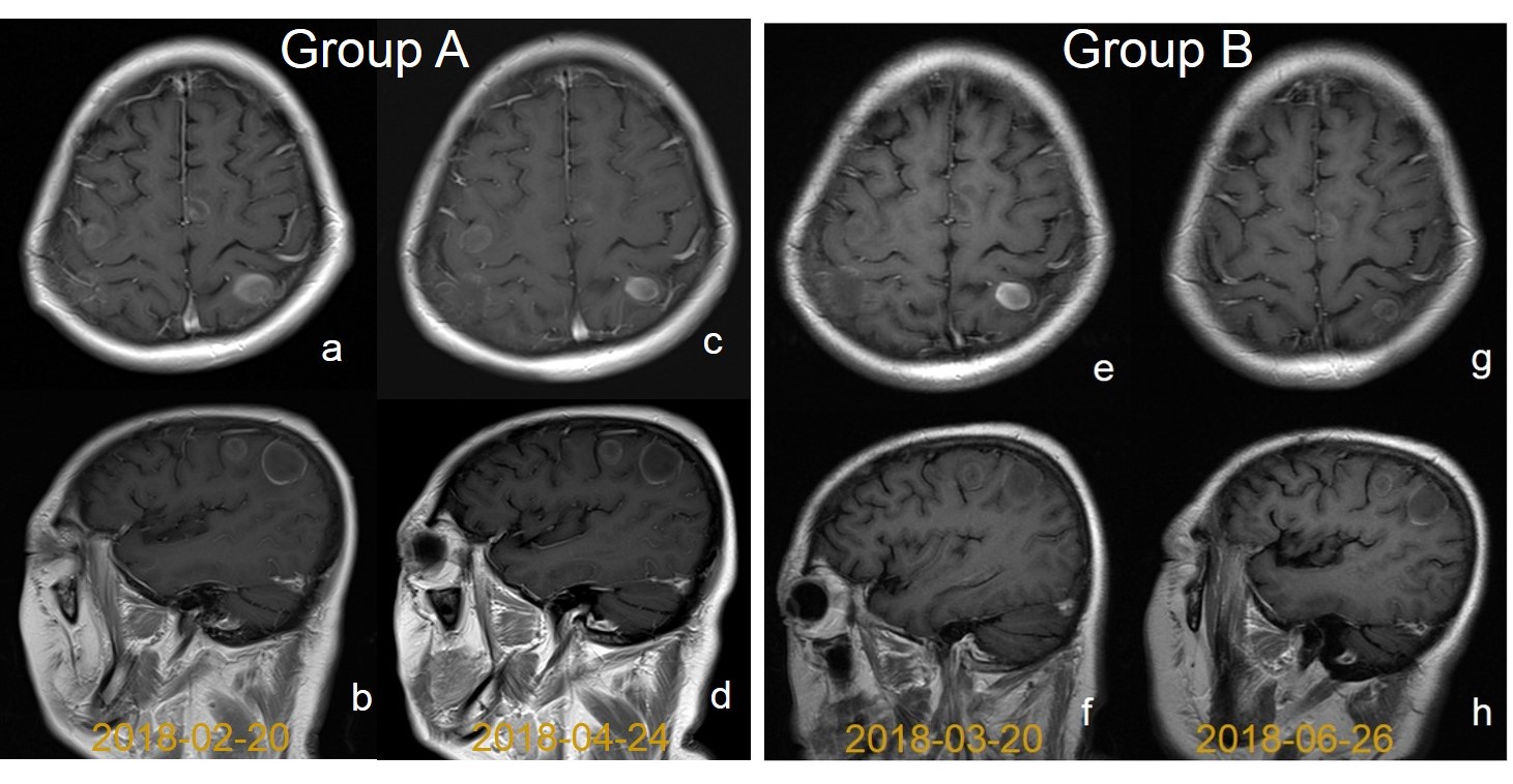
Figure1. Comparison of imagings between groups A and B. In a 66-year-old female with metastatic brain tumors from lung cancer, images from Group A with automatic positioning result in more consistent scanning during follow-up sessions with exactly the same slice positioning.