4753
Identification of the dural tail sign of meningiomas with contrast-enhanced zero echo time (ceZTE) imaging
Lian Jian1, Weiyin Vivian Liu2, Xiaohuang Yang 1, and Xiaoping Yu1
1Radiology, Hunan Cancer Hospital, Hunan, China, 2MR Research, GE Healthcare, Beijin, China
1Radiology, Hunan Cancer Hospital, Hunan, China, 2MR Research, GE Healthcare, Beijin, China
Synopsis
The dural tail sign is often the pathological presence of meningiomas and recently reckoned as a response to vascular congestion and edema. Zero echo time (ZTE) imaging is capable of capturing signal of tissues with relatively short T2* such as pia mater right after onset of the radiofrequency pulse. The significant enhanced rim of meningioma on ecZTE showed the meningioma were supplied by both intracranial and extracranial arteries. Contrast-enhanced ZTE might be listed as a routine imaging sequence to improve accuracy of discriminating the tumor-cortex-dura interface and offer additional clinical information such as vascular growth and supply in meningiomas.
Introduction
The dural tail sign is often a pathological presence in meningiomas and recently reckoned as a response to vascular congestion and edema. It consists merely of a tapering rim rather than a thick extension of the tumor. To discriminate the surface characteristics of dural tail sign is important for a surgery plan due to excision extension depend on its smooth or rough surface.1 Routine MR T1-post-contrast images were applied to delineate the dural tail sign, but convectional T1-post-contrast images is somehow obscure so as not to discriminate pia mater from cortical membrane. In addition, blood supply is another dominant factor to a surgery plan with or without ligation of the carotid artery during operation.2 Therefore, to clearly discriminate extension of dural tail and vascular distribution is of value for clinical surgery planning. A novel zero echo time (ZTE) imaging sequence is capable of capture T2* signal right after onset of the radiofrequency pulse so as to demonstrate tissues with relatively short T2* value on MR images. 3 Therefore, this study aims to explore intracranial meningiomas and pathological conditions with contrast-enhanced ZTE (ceZTE), evaluate the usefulness of ceZTE in detection of the dural tail sign in the meningiomas and compare the performance of ceZTE and contrast-enhanced T1WI (ceT1Wi) on meningiomas.Materials and Methods
This study was approved by the Institutional Review Board of our hospital and all recruited subjects gave the informed consent. 15 patients with meningiomas underwent a routine imaging protocol, including axial T1WI, axial contrast-enhanced T1WI (ceT1WI), and ZTE and contrast-enhanced ZTE (ceZTE), before they were going to receive tumor excision in our hospital from June 2018 to September 2019. 5 cases of meningioma volume based on ceZTE and ceT1WI were obtained with a semi-automatic segmentation tool, ITK-SNAP to perform the quantitative comparison.Results and Discussion
ceT1WI and ceZTE of meningioma and corresponding 3D segmented meningioma were shown in Figure 1. There was no volume difference of meningioma between ceT1WI-based and ceZTE-based segmentation. 15 cases with meningioma showed significant rim enhancement on both ceT1WI, plain ZTE and ceZTE MRI. However, 8 cases of all patients with meningiomas showed more obvious rim enhancement on ceZTE while 4 cases on the plain ZTE sequence or the ceT1WI. The surface of the meningiomas with hyperintensity compared to surrounding tissues might indicated that meningiomas had higher microvascular density supplied by pia mater, indicating ceZTE might be a useful tool to observe vascular growth and distribution as well as blood supply. 14 cases of all showed meningeal tail sign, but10 cases showed greater expression on ceZTE images while 4 cases on ceT1WI.Conclusions
The significant enhanced rim of meningioma on ceZTE demonstrated the meningioma are supplied by both intracranial and extracranial arteries. ceZTE provides pathologically information about the tumor-cortex-dura interface and surface of dura tail sign to make a surgery plan for tumor excision. ceZTE could play an important role in detection of the dural sign of intracranial meningiomas, and thus to make it as a routine imaging sequence might improve accuracy of picturing the tumor-cortex-dura interface and offer additional clinical information such as vascular growth, and distribution as well as blood supply in meningiomas.Acknowledgements
No acknowledgement found.References
- Qi ST, Liu Y, Pan J, et al. A radiopathological classification of dural tail sign of meningiomas. J Neurosurg, 2012, 117(4): 645-653.
- Enokizono M, Morikawa M, Matsuo T, et al. The rim pattern of meningioma on 3D FLAIR imaging: correlation with tumor-brain adhesion and histological grading. Magn Reson Med Sci, 2014, 13(4): 251-260.
- Breighner RE, Endo Y, Konin GP, et al. Zero echo Time imaging of the shoulder: Enhanced Osseous Detail by Using MR Imaging. Radiology, 2018
Figures
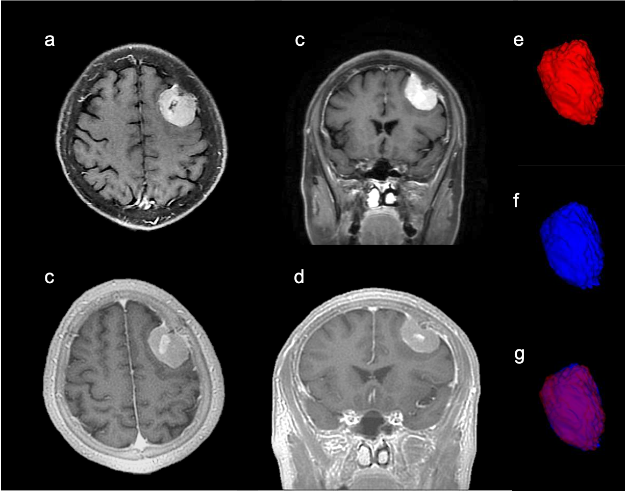
Figure 1(a, b) axial and (c, d)coronal view of ceT1WI and ceZTE, respectively, of 67-year-old female with left meningiomas in the left frontal lobe. The tail sign on ceZTE was more obvious than on ceT1WI. (e, f)3D semi-automatic segmented meningiomas volume based on ceT1WI and ceZTE, respectively. (g)Fusion of the aforementioned segmented volumes. The ceZTE-based segmented volume was bigger than the ceT1W1-based without statistical difference due to small sample sizes (n=5).