4724
Brain and plasma choline compounds altered by cancer and cancer-induced cachexia
Santosh K Bharti1, Paul T Winnard1, Raj Kumar Sharma1, Marie-France Penet1,2, Balaji Krishnamachary1, and Zaver M. Bhujwalla1,2,3
1Div. of Cancer Imaging Research, The Russell H. Morgan Dept of Radiology and Radiological Science, The Johns Hopkins University School of Medicine,, Baltimore, MD, United States, 2Sidney Kimmel Comprehensive Cancer Center, The Johns Hopkins University School of Medicine, Baltimore, MD, United States, 33Radiation Oncology and Molecular Radiation Sciences, The Johns Hopkins University School of Medicine, Baltimore, MD, United States
1Div. of Cancer Imaging Research, The Russell H. Morgan Dept of Radiology and Radiological Science, The Johns Hopkins University School of Medicine,, Baltimore, MD, United States, 2Sidney Kimmel Comprehensive Cancer Center, The Johns Hopkins University School of Medicine, Baltimore, MD, United States, 33Radiation Oncology and Molecular Radiation Sciences, The Johns Hopkins University School of Medicine, Baltimore, MD, United States
Synopsis
Cancer induced cachexia is a multifactorial syndrome that results in unexplained weight loss in cancer patients. A major cause of morbidity and mortality, the syndrome is most prevalent in pancreatic cancer. Here we have identified changes in brain choline metabolites and plasma choline in human pancreatic cancer xenografts that induce cachexia (Pa04C) as well as noncachexia inducing pancreatic cancer xenografts (Panc1), compared to normal mice. These results highlight the systemic changes in choline metabolism that occur with cancer and with cancer induced cachexia that may lead to the development of early biomarkers as well to metabolic treatment strategies.
Introduction
Cancer-induced cachexia is an under explored problem with lethal consequences. To date specialized nutritional supplements have failed as anti-cachexia treatments. There are no known cures for this condition, and the multi-faceted nature of cachexia makes it difficult to investigate. Most prevalent in pancreatic cancer where resection is not possible for a majority of patients, palliation with chemotherapy is the only option of prolonging life. Cachexia results in lower tolerance to chemotherapy [1-3]. Cachectic patients experience a wide range of symptoms affecting the function of organs such as muscle, liver, brain, and heart, causing significant morbidity [4]. An altered choline metabolism is one of the hallmarks of cancer. Changes in choline metabolism in other organs induced by cancers have not been previously investigated. Here, for the first time, we have characterized changes in brain choline metabolism induced by pancreatic cancer xenografts and related these changes to plasma choline levels.Methods
Cancer-induced cachexia is an under explored problem with lethal consequences. To date specialized nutritional supplements have failed as anti-cachexia treatments. There are no known cures for this condition, and the multi-faceted nature of cachexia makes it difficult to investigate. Most prevalent in pancreatic cancer where resection is not possible for a majority of patients, palliation with chemotherapy is the only option of prolonging life. Cachexia results in lower tolerance to chemotherapy [1-3]. Cachectic patients experience a wide range of symptoms affecting the function of organs such as muscle, liver, brain, and heart, causing significant morbidity [4]. An altered choline metabolism is one of the hallmarks of cancer. Changes in choline metabolism in other organs induced by cancers have not been previously investigated. Here, for the first time, we have characterized changes in brain choline metabolism induced by pancreatic cancer xenografts and related these changes to plasma choline levels.Results and Discussion
Cachexia-inducing Pa04C tumors induced a significant weight loss in mice compared to Panc1 tumor bearing mice or normal mice as shown in Figure 1. Representative spectra showing the choline metabolite region of high resolution 1H MRS spectra of brain extracts are shown in Figure 2A. Quantitative analyses of choline, phosphocholine (PC) and glycerophosphocholine (GPC) are summarized in Figures 2B, C and D respectively. A significant decrease of brain choline was observed in Pa04C tumor bearing mice compared to control mice as well as to Panc1 tumor bearing mice, whereas brain PC levels were significantly lower compared to normal mice. Brain GPC was significantly lower in both tumor bearing groups compared to normal mice. We observed a profound decrease of plasma choline in Pa04C tumor bearing mice as shown in Figure 3. Plasma choline in Panc1 tumor bearing mice also decreased but not as drastically. The changes in plasma choline levels most likely contributed to the reduction of brain choline, PC and GPC. Causes of the decrease of plasma choline levels are currently under investigation. Disturbances in the choline/cholinergic pathways may be one underlying cause of the morbidity associated with cancer and cancer-induced cachexia. Reduced plasma choline may represent a risk factor for cancer as choline deficiency is known to result in lymphocyte apoptosis and DNA damage [6] that may compromise the immune response. Normalization of plasma choline using metabolic supplements merits investigation in reducing cancer morbidity and the onset of cachexia.Acknowledgements
This work was supported by NIH R35CA209960 and R01CA193365.References
1. Fearon KC, Baracos VE. Cachexia in pancreatic cancer: new treatment options and measures of success. HPB (Oxford). 2010;12(5):323-4. Epub 2010/07/02. doi: HPB178 [pii] 10.1111/j.1477-2574.2010.00178.x. PubMed PMID: 20590907; PubMed Central PMCID: PMC2951820. 2. Ozola Zalite I, Zykus R, Francisco Gonzalez M, Saygili F, Pukitis A, Gaujoux S, Charnley RM, Lyadov V. Influence of cachexia and sarcopenia on survival in pancreatic ductal adenocarcinoma: A systematic review. Pancreatology : official journal of the International Association of Pancreatology. 2015;15(1):19-24. doi: 10.1016/j.pan.2014.11.006. PubMed PMID: 25524484. 3. Argiles JM, Busquets S, Stemmler B, Lopez-Soriano FJ. Cancer cachexia: understanding the molecular basis. Nature reviews Cancer. 2014;14(11):754-62. doi: 10.1038/nrc3829. PubMed PMID: 25291291. 4. Inui A. Cancer anorexia-cachexia syndrome: current issues in research and management. CA Cancer J Clin. 2002;52(2):72-91. PubMed PMID: 11929007. 5. Winnard PT, Jr., Bharti SK, Penet MF, Marik R, Mironchik Y, Wildes F, Maitra A, Bhujwalla ZM. Detection of Pancreatic Cancer-Induced Cachexia Using a Fluorescent Myoblast Reporter System and Analysis of Metabolite Abundance. Cancer Res. 2016;76(6):1441-50. doi: 10.1158/0008-5472.CAN-15-1740. PubMed PMID: 26719527; PubMed Central PMCID: PMCPMC4794402. 6. da Costa KA, Niculescu MD, Craciunescu CN, Fischer LM, Zeisel SH. Choline deficiency increases lymphocyte apoptosis and DNA damage in humans. The American journal of clinical nutrition. 2006;84(1):88-94. doi: 10.1093/ajcn/84.1.88. PubMed PMID: 16825685; PubMed Central PMCID: PMC2430662.Figures
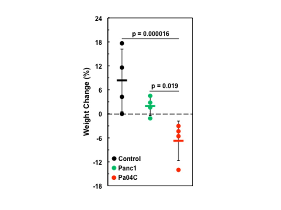
Figure 1: Body
weight changes in normal Control mice as compared to tumor bearing mice. In each cohort several brain weights were
identical, which generated concentric circular representations of brains at the
same weight. Error bars designate standard error of the mean. *Denotes statistical
significance at p < 0.05.
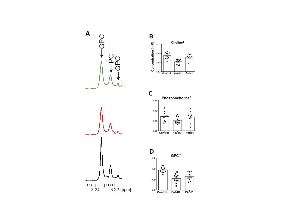
Figure 2: (A) 1H MR spectra of the choline region obtained from aqueous-phase extracts
of brain tissue harvested from non-cachectic Panc1 (top) and cachetic Pa04C
(middle) tumor-bearing mice and normal mice (bottom). GPC: glycerophosphocholine,
PC: phosphocholine, Cho: choline. Changes in (B)
choline (C) PC and (D) GPC in brains of normal mice, mice with cachexia
inducing Pa04C tumors, and mice with non-cachexia inducing Panc1 tumors. Values represent Mean + SEM. Statistical significance was p ≤ 0.06 for: *
Pa04C vs Control, ** Panc1 or Pa04C vs Control, ‡ Pa04C vs Control and Panc1.
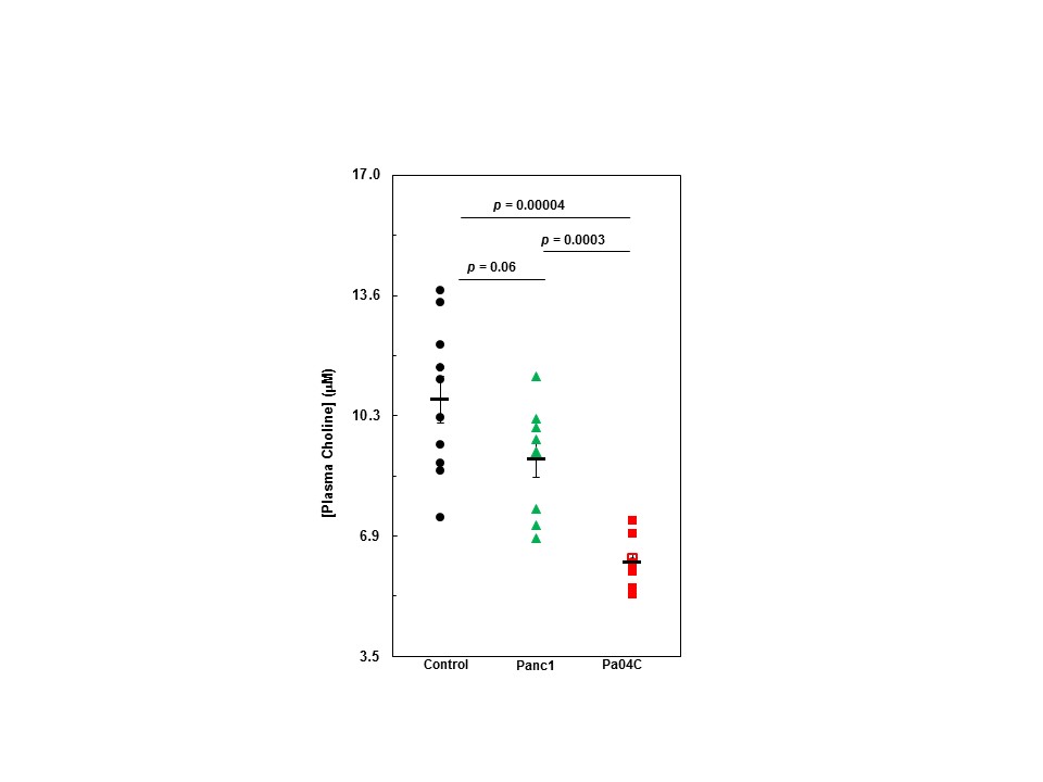
Figure 3: Quantitative
values of mouse plasma choline levels measured using a fluorometric assay. Values represent Mean + SEM.