4706
A Rabbit Model of Intracranial Atherosclerotic Disease for Pathologic Validation of Vessel Wall MRI1University of Utah, Salt Lake City, UT, United States, 2Shengjing Hospital of China Medical University, Shenyang, China, 3Radiology and Imaging Sciences, University of Utah, Salt Lake City, UT, United States
Synopsis
Two rabbit model types (Watanabe Heritable Hyperlipidemic and Apolipoprotien E knockout) were studied with vessel wall MRI (vwMRI) and confirmatory histopathological analysis to evaluate for presence of intracranial atherosclerosis (ICAD). Disease was reliably produced in both rabbit types. Sensitivity and specificity of vwMRI for detection of ICAD were 89.5% and 91.9%, respectively. When excluding mild cases to assess accuracy vwMRI for detecting moderate to severe ICAD lesions, sensitivity improved to 100% with unchanged specificity.
Purpose
Vessel wall MRI (vwMRI) sequences have improved characterization of vascular disorders by allowing visualization of the vessel walls themselves, adding to prior information obtained from older techniques that only imaged the vessel lumen only. AmonKOg the disorders evaluated by vwMRI, intracranial atherosclerotic disease (ICAD) in particular can now be more clearly visualized by imaging the vessel walls. ICAD is a leading cause of ischemic stroke, causing long-term morbidity and mortality worldwide. Current controversy in stroke risk reduction treatment approaches results from limited understanding of the pathophysiologic drivers of ICAD. This in part results from a paucity of histopathological data from human lesions, which can typically only be obtained at autopsy. Furthermore, drug discovery and mechanistic studies in humans cannot realistically be acquired. Therefore development of a 3T vwMRI-compatible animal model would be extremely useful for preclinical studies. This study seeks to characterize vwMRI findings in Watanabe heritable hyperlipidemic (WHHL) and apolipoprotein E-knockout (ApoE-/-) rabbits with comparison to histopathological results.Methods
WHHL at advanced age underwent serial vwMRI studies on a 3T Prisma scanner (Siemens Healthineers, Erlangen, Germany) using an ankle coil over the head to obtain pre- and post-contrast DANTE T1 SPACE, T2 SPACE, and TOF MRA. Table 1 summarizes the DANTE preparatory pulse sequence parameters. Mature WHHL animals demonstrated advanced disease, but during the course of the study, younger WHHL rabbits became unavailable due to logistical limitations at the breeding facility. To evaluate earlier disease, mature ApoE rabbits fed a high cholesterol diet for up to six months underwent the same imaging protocol. Additionally, juvenile wild-type New Zealand White (NZW) rabbits were evaluated according to the same protocol to serve as normal controls. When determined appropriate to evaluate a certain stage of disease or dictated by failure to thrive, euthanasia was performed with perfusion fixation, delivering steady flow of 2% paraformaldehyde and 5% glutaraldehyde solution through a vascular sheath inserted percutaneously into a carotid artery. With perfusate flowing, a jugular vein was transected to exsanguinate the animal while achieving fixation with few blood cells in vessels. The animal was decapitated and the brain and intracranial arteries harvested and placed in a container with formalin. After two weeks, specimens were sliced to prepare slides oriented to maximize the cross-sectional orientation of the proximal intracranial arteries. Light microscopic analysis was performed after hematoxylin and eosin (H&E) staining to assess the presence of ICAD. Slides were analyzed by a specialized veterinary pathologist. vwMRI images obtained closest to euthanasia were analyzed by a board certified radiologist with certificate of added qualification in neuroradiology. Basilar and internal carotid artery segments were each rated as having no, mild, moderate, or advanced ICAD on both microscopic and vwMRI samples. Two-tailed t-tests were performed to compare disease prevalence among rabbit subtypes. Sensitivity and specificity were calculated to assess diagnostic accuracy of vwMRI.Results
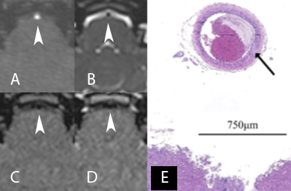
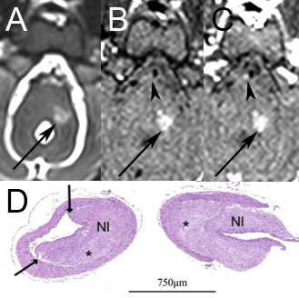
17 rabbits underwent evaluation, including 5 WHHL (24.3-31.6 months at euthanasia, mean 27.4 ± 2.8), 4 ApoE (37.8-46.8 months, mean 42.8 ± 4.2), and 8 NZW rabbits (6.1-20.9 months, mean 11.1 ± 5.2). vwMRI and histopathology images representative of normal vessels and severe disease are provided in Figures 1 and 2, respectively. 5 (33.3%) vessel segments had ICAD identified on pathology (2 mild, 2 moderate, 1 severe) in WHHL animals, while 6 (50.0%) ApoE vessel segments had lesions (5 mild, 1 moderate). No lesions were identified in NZW animals. Two-tailed t-tests confirmed that disease model animals had lesions more frequently than NZW animals (p<0.001). There was no statistical difference in disease occurrence between WHHL and ApoE animals (p=0.178). The observed presence of more advanced disease noted in WHHL rabbits compared to ApoE rabbits was not statistically significant (p=0.296). Sensitivity and specificity of vwMRI for detection of ICAD were 89.5% and 91.9%, respectively. When excluding mild cases to assess vwMRI accuracy for detecting moderate to severe ICAD lesions, sensitivity improved to 100% with unchanged specificity.Discussion
ICAD can be reliably produced and detected using a 3T vwMRI-compatible rabbit model. Mature WHHL rabbits demonstrated ICAD. Mature ApoE rabbits demonstrated less advanced ICAD after short term exposure to a hyperlipidemic diet. Further analysis is needed to better characterize development and progression of disease in ApoE rabbits. vwMRI sequences can be used to analyze the disease in these model animals with high sensitivity and specificity.Acknowledgements
No acknowledgement found.References
1. Ji D, Zhao G, Songstad A, Cui X, Weinstein EJ. Efficient creation of an APOE knockout rabbit. Transgenic Res. 2015 Apr;24(2):227-35. doi: 10.1007/s11248-014-9834-8. Epub 2014 Sep 13.
2. Kong J, Tamaki N, Asada M. Early lesions of cerebral atherosclerosis from induced hypertension in Watanabe heritablehyperlipidemic rabbits. Kobe J Med Sci. 2000 Apr;46(1-2):87-101.
3. Alexander MD, Yuan C, Rutman A, Tirschwell DL, Palagallo G, Gandhi D, Sekhar LN, Mossa-Basha M. High-resolution intracranial vessel wall imaging: imaging beyond the lumen. J Neurol Neurosurg Psychiatry. 2016 Jun;87(6):589-97.
Figures