4687
Associations of body size and composition with regional brain volumes and white matter microstructure in very preterm infants1Department of Pediatric Newborn Medicine, Brigham & Women's Hospital, Boston, MA, United States, 2Harvard Medical School, Boston, MA, United States, 3Computational Radiology Laboratory, Department of Radiology, Boston Children's Hospital, Boston, MA, United States, 4Department of Pediatric Surgery, Research Field in Medical and Health Sciences, Medical and Dental Area, Research and Education Assembly, Kagoshima University, Kagoshima, Japan
Synopsis
For very preterm infants, body size and composition (lean versus fat mass) may index brain growth and microstructural development. Among 85 very preterm infants at term equivalent age, we studied associations of body size/composition with brain magnetic resonance imaging (MRI) outcomes including total and regional brain volumes, and fractional anisotropy of white matter tracts. Larger body size and more lean--but not fat--mass were associated with larger brain volumes and higher fractional anisotropy of multiple white matter tracts. Lean mass accrual may index brain growth and development. MRI may be useful for studying effects of nutritional exposures on the preterm brain.
Introduction
For very preterm infants, birth to term equivalent age is a critical period for brain growth and development. Differentiating lean from fat mass provides information about the quality of an infant’s overall physical growth during this period and may also index brain growth and maturation.1–3 Specific regions of the brain (hippocampus, cerebellum) and developmental processes (neuronal proliferation and myelin production) are particularly sensitive to nutritional perturbations4, but little is known about the relationships between body composition and regional brain growth or white matter maturation. Therefore, our objective was to assess associations of anthropometric size and body composition (lean and fat mass) with (1) regional brain volumes and (2) white matter microstructure at term equivalent age among very preterm infants. We hypothesized that greater lean mass—but not fat—would be associated with larger brain volume, particularly in the hippocampus and cerebellum, and with higher fractional anisotropy of early myelinating white matter tracts.Methods
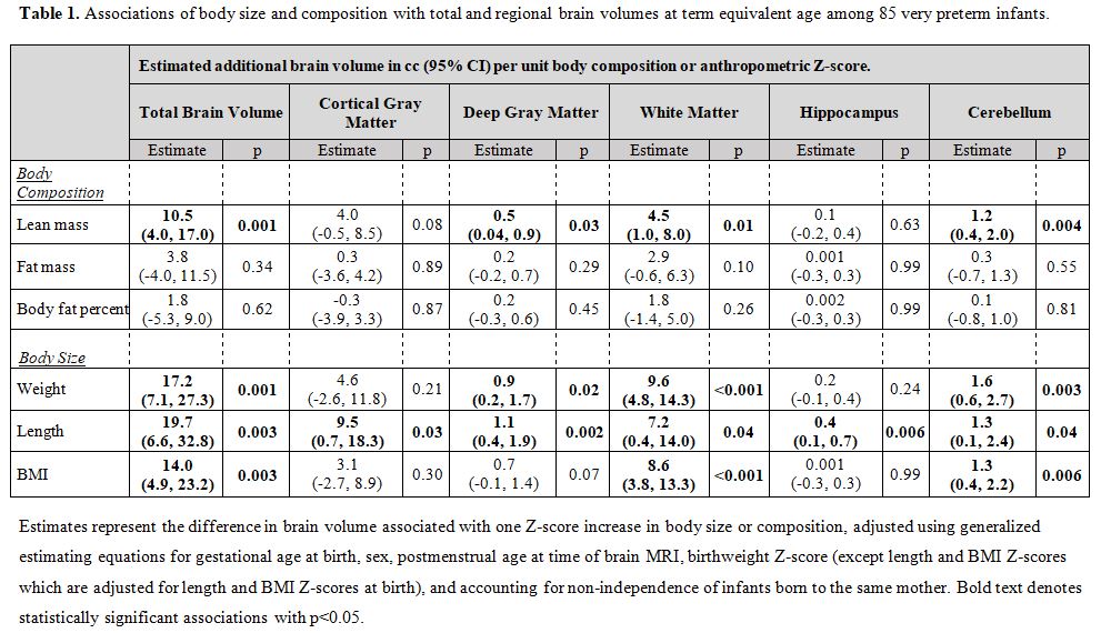
We enrolled 85 infants born <33 weeks’ gestation in a prospective observational study. At median postmenstrual age 39.3 weeks, infants underwent body composition measurement using air displacement plethysmography and brain magnetic resonance imaging (MRI) using a Siemens Trio 3 Tesla scanner (Erlangen, Germany). T2-weighted images were acquired with a sagittal T2 turbo spin echo sequence, 1 mm isotropic voxels, flip angle = 160°, TR = 8630 ms, TE = 133 ms, FOV = 190 x 190 mm, matrix = 192 x 192. Diffusion images were acquired with 30 directions at b=1000s/m2 with 1 b=0, and 2 mm isotropic voxels. We used automated segmentation (MANTiS)5 to generate volumes of the cortical gray matter, deep gray matter, white matter, hippocampus, cerebellum, and total brain. Diffusion tensor imaging with an automated tractography framework6 was used to generate fractional anisotropy (FA) and mean, radial, and axial diffusivity of 15 white matter tracts including the corpus callosum, and bilateral anterior thalamic radiations, cingulum, corticospinal tracts (CST), inferior longitudinal fasciculi, optic radiations, posterior limb of the internal capsule (PLIC), and uncinate fasciculi. From body composition measurements, we calculated Z-scores of lean and fat mass using reference values for infants born full term7. We determined Z-scores of weight, length, and body mass index (BMI) at term equivalent age using the Olsen reference charts8,9. We estimated cross-sectional associations of body size and composition with brain volumes and diffusion measures in models adjusted for gestational age at birth, sex, birthweight Z-score (as a proxy for fetal growth), postmenstrual age at time of MRI, and using generalized estimating equations to account for non-independence of multiple births.Results
Participants were 57% male, with median gestational age 29.1 weeks (range 23.4, 32.9). Anthropometric size, including weight, length, and BMI, was positively associated with volumes of most brain regions and total brain volume (Table 1). Greater lean mass at term equivalent age was associated with larger volumes of most brain regions (Table 1). Specifically, in adjusted analyses, each unit higher lean mass Z-score was associated with significantly greater mean brain volumes as follows: total brain 10.5cc (95% confidence interval [CI]: 4.0, 17.0), deep gray matter 0.5cc (95%CI: 0.04, 0.9), white matter 4.5cc (95%CI: 1.0, 8.0), and cerebellum 1.2cc (95%CI: 0.4, 2.0). With respect to diffusion outcomes, anthropometric size and lean mass were positively associated with FA in multiple white matter tracts (Table 2). Specifically, a one unit increase in lean mass Z-score was associated with greater FA as follows: left cingulum 0.3%; left corticospinal tract 0.5%; and right PLIC 0.3%. There were no associations of lean mass with FA in the remaining tracts (Table 2). There were also no associations between any exposure variable and mean, radial, or axial diffusivity (data not shown). In contrast to lean mass, associations between fat Z-scores and brain volumes or diffusion measures had smaller effect sizes and were not statistically significant (Tables 1 and 2).Discussion/Conclusions
Greater lean mass, but not fat, at term equivalent age was associated with larger volume of most brain regions and total brain size, as well as microstructural alterations, namely greater FA in multiple white matter tracts. Lean mass accrual may index brain growth among preterm infants as well as white matter microstructural changes. Nutritional factors that promote greater lean mass accretion may also promote increased brain growth and white matter maturation. Brain MRI may be a useful tool to elucidate effects of nutritional exposures on the developing preterm brain.Acknowledgements
No acknowledgement found.References
1. Bell KA, Matthews LG, Cherkerzian S, et al. Associations of Growth and Body Composition with Brain Size in Preterm Infants. The Journal of Pediatrics. 2019;214:20-26.e2.
2. Ramel SE, Gray HL, Christiansen E, et al. Greater Early Gains in Fat-Free Mass, but Not Fat Mass, Are Associated with Improved Neurodevelopment at 1 Year Corrected Age for Prematurity in Very Low Birth Weight Preterm Infants. J Pediatr. 2016;173:108-115.
3. Coviello C, Keunen K, Kersbergen KJ, et al. Effects of early nutrition and growth on brain volumes, white matter microstructure, and neurodevelopmental outcome in preterm newborns. Pediatric Research. 2018;83(1-1):102-110.
4. Keunen K, van Elburg RM, van Bel F, Benders MJNL. Impact of nutrition on brain development and its neuroprotective implications following preterm birth. Pediatr Res. 2015;77(1-2):148-155.
5. Beare RJ, Chen J, Kelly CE, et al. Neonatal Brain Tissue Classification with Morphological Adaptation and Unified Segmentation. Front Neuroinform. 2016;10:12.
6. Suarez RO, Commowick O, Prabhu SP, Warfield SK. Automated delineation of white matter fiber tracts with a multiple region-of-interest approach. Neuroimage. 2012;59(4):3690-3700.
7. Norris T, Ramel SE, Catalano P, et al. New charts for the assessment of body composition, according to air-displacement plethysmography, at birth and across the first 6 mo of life. The American Journal of Clinical Nutrition. 2019;109(5):1353-1360.
8. Olsen IE, Groveman SA, Lawson ML, et al. New intrauterine growth curves based on United States data. Pediatrics. 2010;125(2):e214-224.
9. Olsen IE, Lawson ML, Ferguson AN, et al. BMI curves for preterm infants. Pediatrics. 2015;135(3):e572-581.
Figures