4679
Pediatric acute mTBI study of GABA concentration in posterior cingulate cortex1Emanuel Institute of Biochemical Physics of the Russian Academy of Sciences, Moscow, Russian Federation, 2Clinical and Research Institute of Emergency Surgery and Trauma, Moscow, Russian Federation, 3Clinical and Research Institute of Emergency Pediatric Surgery and Traumatology, Moscow, Russian Federation, 4Semenov Institute of Chemical Physics of the Russian Academy of Science, Moscow, Russian Federation
Synopsis
This is the first measurement of pure GABA concentration in the PCC of children with mild TBI in the acute phase. MEGA-PRESS pulse sequence was used in order to obtain GABA signal not contaminated by macromolecules. The result obtained disagrees with the previous study, where [GABA] was increased in the anterior cingulate cortex of children with mTBI when comparing to healthy controls. The findings may signify that ACC is more sensitive to mTBI than PCC.
Introduction
The mechanisms underlying functional disorders in the central nervous system caused by traumatic brain injury require intensive research. In 10–-25% of mTBI patients, post-concussion symptoms persist over several month or longer, significantly impairing the quality of life. One possible reason of posttraumatic disorders could be the imbalance between major neurotransmitters (excitatory – glutamate, and inhibitory – γ-aminobutyric acid, GABA), that is crucial for the normal functioning of CNS. In this study, [GABA] is measured in the posterior cingulate cortex (PCC) of children with acute mTBI.Materials & Methods
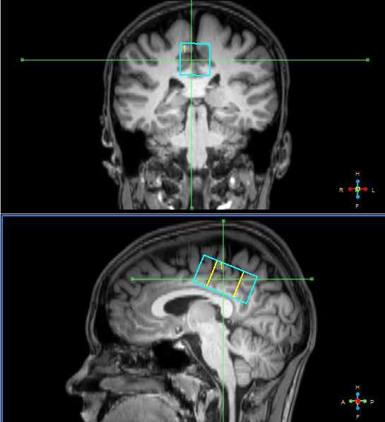
Twelve patients with acute mTBI (12–70 hours since the injury, aged 15.7±1.9) and thirteen healthy subjects (aged 19.3±0.7) participated in the study. MRI scanner Philips Achieva dStream 3.0T was used. Standard MRI protocol for TBI patients (T2-, T1-weighted images, FLAIR, SWI, DTI) revealed no pathological lesions in brain tissue of any subject were found. Magnetic resonance spectroscopy voxels were located in the area of posterior cingulate cortex (see figure 1). MEGA-PRESS pulse sequence was used in order to obtain GABA signal not contaminated by macromolecules [1]: TR=2000 ms, TE=80 ms, 20 ms editing pulses on δOn=1.9 ppm, δOff=1.5 ppm, NSA=288, acquisition time ~10 min. TE averaged PRESS spectrum [2] was acquired to obtain Glu separated from glutamine: TR = 2000 ms, TE starting from 35 ms up to 185 ms with the 2.5 ms step, NSA for each TE=4, (acq time ~4.5 min). After that, standard PRESS spectrum (TE=35 ms, TR=2000 ms, NSA=128) and additional water reference spectrum (TE=28 ms, TR=10000 ms, NSA=4) were obtained in the same voxel. GABA spectra were processed in Gannet 3.0 [3], standard PRESS spectra and TE averaged PRESS – in LCModel. For basis set, TE averaged PRESS spectra of cerebral metabolites were simulated in FID-A. Absolute concentrations of GABA and conventional MRS-detectible metabolites were calculated, taking into account voxel grey/white/CSF composition as well as correction for the relaxation effects. Normalized on Cr values were analyzed as well. Statistical analysis was performed in STATISTICA 12. The Mann-Whitney criterion was used to reveal the significance of between-group differences.Results
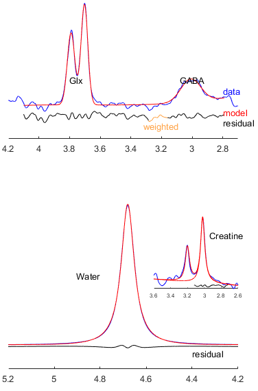
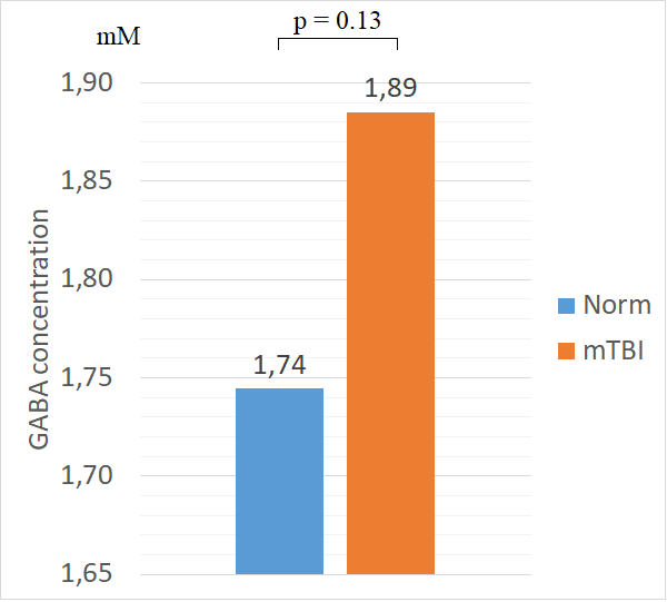
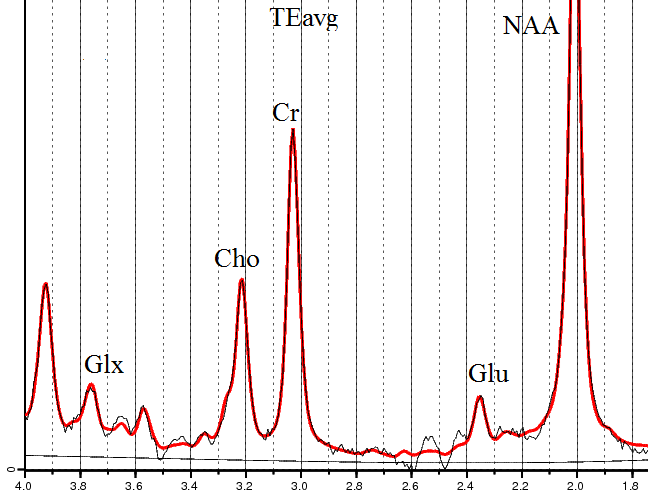
Typical GABA spectrum processing in Gannet is demonstrated on figure 2. The values of [GABA] in PCC are demonstrated on figure 3: the increase in GABA in PCC in mTBI group is not statistically significant. No between-group difference in GABA/Cr and GABA/Glx was found as well. Typical TEavg spectrum processing is shown on figure 4. Glu/Cr values in the normal and mTBI groups, found from TE averaged PRESS, were equal. The absolute concentrations of other standard MRS-detectible metabolites also demonstrated no between-group differences.Conclusion
To our knowledge, this is the first measurement of pure GABA concentration in the PCC of children with mild TBI in the acute phase. The result obtained in current work is not in agreement with the result of our previous study, where [GABA] was increased (p<0.005) in the anterior cingulate cortex (ACC) of children with mTBI when comparing to healthy controls [4]. Unlike [4], GABA/Glx values in this work are also not elevated in mTBI group. These findings may signify that ACC is more sensitive to this type of injury than PCC. They also indicate a necessity of further data collecting (e.g. in different regions) in order to discover [GABA] alterations in various cerebral loci. This would help to identify the cases of an inhibition/excitation imbalance and to predict possible dysfunctions of the CNS following mild TBI.Acknowledgements
We thank Prof. Peter Barker and Prof. Richard Edden from Johns Hopkins University for the HERMES patch that allows to modify the MEGA-PRESS pulse sequence.
The work is supported by the RFBR grant 18-315-00165.
References
[1] 10.1002/mrm.24391
[2] 10.1002/mrm.20007
[3] 10.1002/jmri.24478
[4] 10.1134/S0006350917060161
Figures