4675
Spatiotemporal changes of optic radiation from 3-13 years: a diffusion tensor imaging study1the First Affiliated Hospital of Xi’an Jiaotong University, Xi’an, China, 2MR Research China, GE Healthcare, Xi’an, China
Synopsis
Optic radiations(OR), which connecting the lateral geniculate nuclei and visual cortex, plays a critical role in visual function. Detailing the maturation process of OR is helpful in understanding the visual development and identifying the abnormalities. Diffusion tensor imaging can quantify the brain white matter development. Using DTI-based automating fiber-tract quantification, this study explored the spatiotemporal OR changes in children aged 3-13 years. Results indicated that the anterior, middle, and posterior segments of OR showed asynchronous developmental patterns. Specifically, the mid-segment of OR presented more mature than the anterior segment, while right OR showed more extended mature than left OR.
Introduction
Visual function development is closely related to the fulfillment and myelination of brain white matter (WM) connections1. Optic radiation (OR), which connecting lateral geniculate nuclei (LGN) and visual cortex, launches its myelination in 3 to 4 months of life and reaches the adult level by 3 years of age2,3. Previous studies found the developmental changes of the neonatal optic radiation microstructure. But few studies involved the OR development in children. Given the critical period of visual function, this study aims to explore the spatiotemporal changes of OR from 3-13 years and lay the foundation for further study of the maturity of visual function.Methods
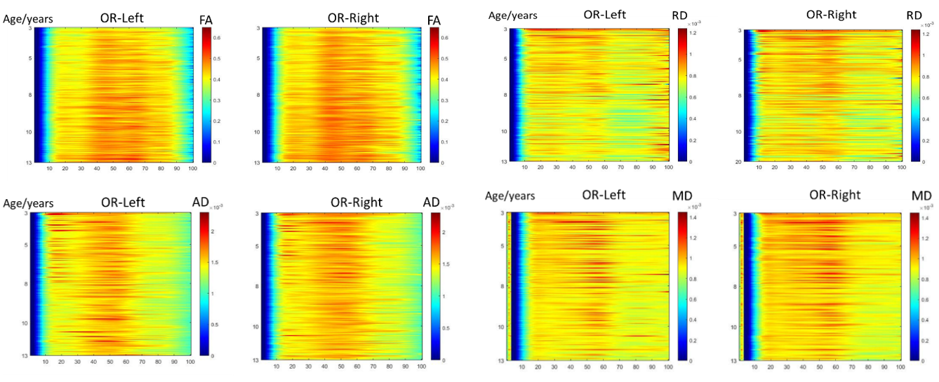
This study was approved by the local Internal Review Board and all parents of participants had signed the informed consents before MRI scanning. Subjects A total of 154 children with diffusion tensor imaging (DTI) scanning aged 3-13 years (111 boys, 43 girls) were retrospectively collected and all born at full-term without abnormalities in brain MR images. MR Protocols All MR examinations were performed using a 3.0T scanner (Signa HDxt, GE Healthcare, Milwaukee, Wisconsin) with an 8-channel head coil. The parameters of DTI sequence were as follows: 35 gradient directions; b-values =0 and 1000 s/mm2; TR/TE= 5500/95–105 ms; section thickness=4mm without a gap; FOV=180 × 180 mm2; matrix size =128 × 128; and voxel size= 1.41 × 1.41 × 4 mm3. Data and statistical analysis DTI data were preprocessed by FMRIB software library (FSL, http://www.fmrib.ox.ac.uk/fsl) and Matlab software (MathWorks, Natick, Massachusetts). Quantitative tracking of optic radiation was performed using Automating Fiber-Tract Quantification (AFQ). Four parameters of fractional anisotropy(FA), mean diffusivity(MD), axial diffusivity(AD),and radial diffusivity(RD) were calculated by using the above tools. The OR is equally divided into 100 sites, and the average value of the vertical section DTI parameters of each locus is calculated(The 1-34th sites, the anterior segment; the 35th-67th sites, the middle segment; the 68th-100th sites, the posterior segment). Spearman and Pearson correlation analysis were used to analyze the correlation between DTI parameters and age and vision. All statistical analysis was performed by using SPSS 18.0(SPSS, Chicago, IL,USA); p<0.05 was considered as statistically significant difference.Results
In this study, the mean values of FA, RD, AD, and MD of optic radiation were correlated with age coefficient of 0.578(P<0.05), -0.361(P<0.05), -0.293(P<0.05) and -0.471(P<0.05). In the colormaps, optic radiations exhibit different spatial developmental features especially in FA and MD values. The FA value of the middle segment is higher than that in the anterior and posterior segments. And the posterior segment showed a significant decrease in MD and AD values (Figure 1).Discussion
By using DTI, this study quantified the spatiotemporal changes of OR microstructure in normally developing children. We found that the anterior, middle, and posterior segments of OR showed asynchronous developmental patterns. Previous studies found the developmental changes of the optic radiation microstructure of neonates. In neonates, the anterior segment of OR matures earlier than the posterior segment4. And based on a healthy population research aged 5-18 years old, there was no obvious age effect on the volume of OR, but both suggested FA was increased by age5. Our results suggested OR segments may have different maturation process. Although the mid-segment of myelination develops later than the anterior segment, the mid-segment maturity may gradually earlier by age.Conclusion
Being different from neonates, OR showed an asynchronous developmental patterns from 3-13 years. Specifically, the mid-segment presented more mature than the anterior segment, while right OR showed more extended mature than left OR.Acknowledgements
This study was supported by the National Natural Science Foundation of China (81901516, 81971581, 81901823, 81771810 and 51706178), the 2011 New Century Excellent Talent Support Plan of the Ministry of Education, China (NCET-11-0438) and the Clinical Research Award of the First Affiliated Hospital of Xi’an Jiaotong University (No. XJTU1AF-CRF-2015-004).References
1.Dubois, J, Dehaene LG, Kulikova, S, et al. The early development of brain white matter a review of imaging studies in fetuses newborns and infants. Neuroscience, 2014, 276 :( 48–71)
2.Kinney HC1, Brody BA, Kloman AS, et al. Sequence of central nervous system myelination in human infancy. II. Patterns of myelination in autopsied infants. J Neuropathol Exp Neurol,1988, 47(3):217-34.
3.Deoni SC, Mercure E, Blasi A, et al. Mapping infant brain myelination with magnetic resonance imaging. J Neurosci, 2011,31(2):784–91.
4. Dubois J, Dehaene LG, Soares C, et al. Microstructural correlates of infant functional development: example of the visual pathways. Journal of neuroscience, 2008, 28(8): 1943-1948.
5. Dayan M, Munoz M, Jentschke S, et al. Optic radiation structure and anatomy in the normally developing brain determined using diffusion MRI and tractography. Brain structure & function,2015, 220(1): 291-306.
Figures