4670
How does Microstructural in lntra-axonal and Extra-axonl Space Change in Preterm Infants with Punctate White Matter Lesions?1Radiology, the First Affiliated Hospital, Xi'an Jiaotong University, Xi'an, China, Xi'an, China, 2MR Research China, GE Healthcare, Bei Jing, China
Synopsis
Punctate white matter lesions (PWML)are common in preterm infants.Extensive microstructural changes were observed previously for different PWML grades by using diffusion tensor imaging (DTI).However, the changes of intra-axon and extra-axon remain to be investigated. White matter tract integrity (WMTI)metrics derived from diffusion kurtosis imaging (DKI)provide information of intra-axonal or extra-axonal spaces. Our study aimed to use WMTI metrics to detect the Microstructural Variations.Compared to DTI metrics, the change trends of fractional anisotropy (FA),axial diffusivity (AD) and radial diffusivity (RD)are in agreement with previous findings.Furthermore, intra-axonal diffusivity (Da)unchange or reduce in lntra-axonal space.There were increased RDe, reduced/unchanged ADe in extra-axonal space.
Introduction
Punctate white matter lesions (PWML) have been found in more than 20% of preterm infants1, 2. These lesions may cause severe neurologic disorders, such as cerebral palsy2, 3. PWMLs without cystic lesions can be divided into 3 grades4. Severe research have found that PWML grade III could cause extensive changes in white matter microstructure by using diffusion tensor imaging (DTI)5. However, conventional DTI parameters cannot distinguish the development of axons themselves from the growth of myelin6. White matter tract integrity (WMTI) metrics derived from diffusion kurtosis imaging (DKI) can provide information of intra-axonal or extra-axonal spaces. Metrics in the lntra-axonall space include intra-axonal diffusivity (Da). Metrics in the extra-axonal space include extra-axonal radial diffusivity (RDe) and extra-axonal axial diffusivity (ADe). This study aimed to use WMTI metricsto quantify characterize the Microstructural Variations in Intra-axonal and Extra-axonal Spaces with PWML Infants.Methods
This study was approved by the local Internal Review Board and all parents of participants had signed the informed consents. Inclusion criterion was the evidence of punctate lesions in the cerebral white matter, which presented on T1WI and T2WI. Preterm infants without any MRI abnormality were selected as controls. Subjects with clinical diagnosis of congenital malformations of the central nervous system, hydrocephalus, gray matter lesions or major destructive white matter lesions were excluded.Single-shot EPI diffusion kurtosis imaging was performed on a 3.0T scanner (General Electric Signa HDXT, WI, USA) with an eight-channel head coil. The other parameters were: b values = 500, 1000, 2000, 2500 s/mm2; 18 gradient directions; TR/TE = 8000~11000/91.7~126.1 ms; thickness = 4 mm; FOV = 180 × 180 mm2 ~ 240 × 240 mm2 (according to brain sizes); acquisition matrix = 128 × 128 ~ 172 × 172 (to keep the same resolution). Diffusion and kurtosis tensors were estimated by using constrained weighted linear least squares. Fractional anisotropy (FA), axial diffusivity (AD), radial diffusivity (RD), and WMTI metrics were calculated according to the white matter model for DKI.
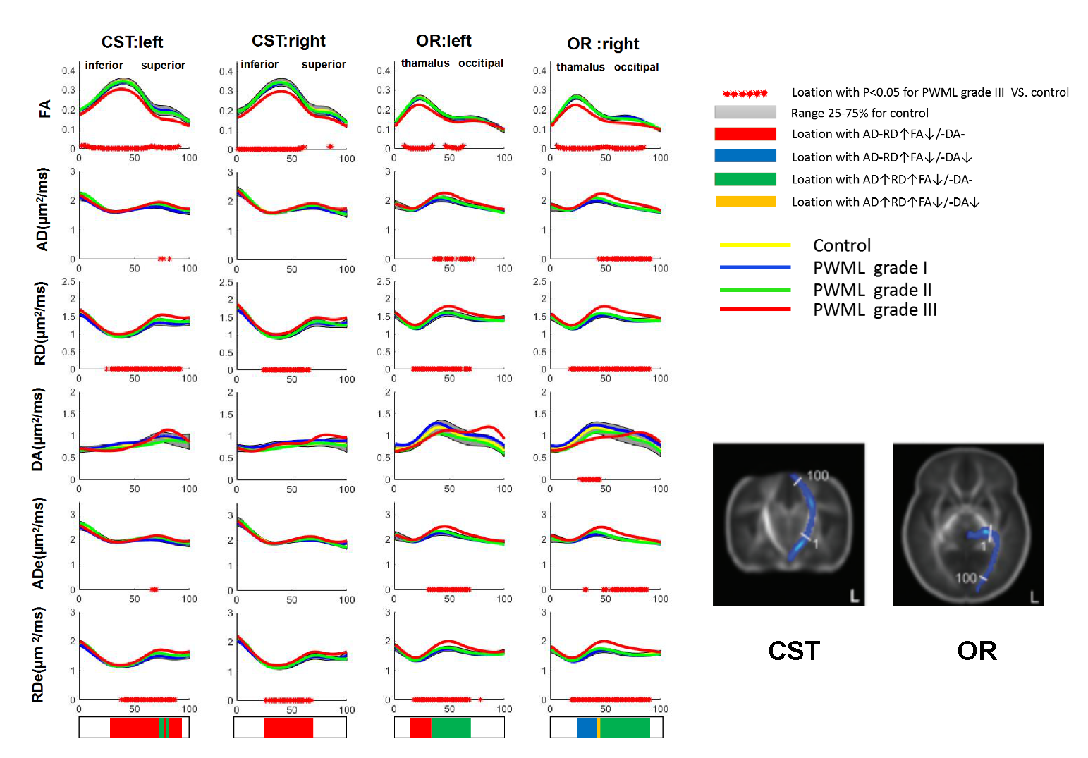
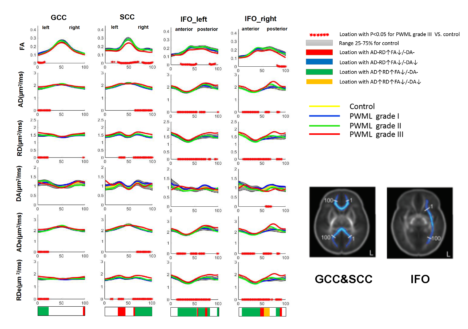
Tract-Quantification Analysis:we quantified the DKI metrics on the representative tracts: projection fibers of the corticospinal tract (CST) and optic radiation (OR); commissural fibers of the splenium of the corpus callosum(SCC)and genu of the corpus callosum (GCC); and association fibers of the inferior fronto-occipital fasciculus (IFO).DTI data were preprocessed by FMRIB software library( FSL; http://www.fmrib.ox.ac.uk/fsl) and Matlab software (MathWorks, Natick, Massachusetts). Quantitative tracking of optic radiation fiber bundles was performed using Automating Fiber-Tract Quantification (AFQ). FA, AD, RD, and WMTI metrics were calculated by using the above tools.
Result
A total of 36PWML and 32control preterm infants were included.19, 7, and 9 preterm infants with PWMLs were classified into grades I, II, and III, respectively. There were no significant differences in gender ratio, gestational age (GA), postnatal age and postmenstrual age (PMA) between PWML and control groups (Table 1).Patients with PWML grade I and grade II had no change in all DKI parameters compared to control preterm infants .Different patterns of microstructural changes associated with severe PWMLs (grade III) were found along white matter tracts. Unchanged AD, increased RD, unchanged Da and reduced/reduced FA were found in the inferior, central and superior parts of the left CST, the central part of the right CST, the occipital and thalamus proximal regions of the left OR,the occipital,central and thalamus proximal regions of the right OR, the left region of GCC, the left and central regions of SCC, the central region of the left IFO. Unchanged AD. Increased RD, reduced Da and reduced/reduced FA were found in the inferior parts of the left CST, the occipital region of the right OR and the central region of the right IFO. Reduced AD, increased RD, unchanged Da and reduced/reduced FA were found in the inferior, central and superior parts of the left CST, ,the central and occipital proximal regions of the right OR, the left region of GCC, the central and right regions of SCC, the anterior, central and posterior regions of the left IFO, the anterior and posterior regions of the right IFO. Reduced AD, increased RD, reduced Da and reduced/reduced FA were found in the posterior regions of the right IFO. Areas and trends of ADe and RDe changes are similar to AD and RD (Figure 1 and
Figure 2).
Disccussion
This study demonstrated extra-axonal and intra-axonal structural changes associated with PWML grade III. Compared to previous DTI studies 1.5. Besides reduced FA and increased RD, increased AD were found. Furthermore, reduced Da, increased RDe and ADe were found.TheVariations of extent and scope for changes are not as obvious as RDe and RDa.previous study has found that extra-axonal metrics are sensitive to myelin-related alterations7. Results here indicate that PWML may cause dysmyelination in infants. The decrease of Da may indicate the edema of axon8. Our study reveal that all the microstructural in intra-axonal and extra-axonl Space Change in Preterm Infants with PWML grade III. And the Variations in extra-axonl space are more significiant than in intra-axonal space.
Conclision
All the microstructural in intra-axonal and extra-axonl Space Change in Preterm Infants with PWML grade III. And the Variations in extra-axonl space are more significiant than in intra-axonal space.Acknowledgements
This study was supported by the National Natural Science Foundation of China (81971581, 81901823, 81771810), National Key Research and Development Program of China (2016YFC0100300), the 2011 New Century Excellent Talent Support Plan of the Ministry of Education of China (NCET-11-0438), the Project Funded by China Postdoctoral Science Foundation (2019M653659), and the Natural Science Basic Research Plan in Shaanxi Province of China (2019JQ-198).
References
1. Bassi L, Chew A, Merchant N, et al. Diffusion tensor imaging in preterm infants with punctate white matterlesions. Pediatr Res 2011;69:561-566.
2. De Bruïne FT, van den Berg-Huysmans AA, Leijser LM, et al. Clinical implications of MR imaging findings in thewhite matter in very preterm infants: a 2-year follow-up study. Radiology 2011;261:899-906.
3. Kersbergen KJ. Different patterns of punctate white matter lesions in serially scanned preterm infants. PLOS One 2014;9:e108904.
4. Childs AM, Cornette L, Ramenghi LA, et al. Magnetic resonance and cranial ultrasound characteristics of periventricular white matter abnormalities in newborn infants. Clin Radiol 2001;56:647–55.
5. Li X, Gao J, Wang M, et al. Charaterization of extensive microstructural variations associated with punctate white matter lesions in preterm neonates. AJNR Am J Neuroradiol 2017;38:1228 -1234.
6. Paus T.Growth of white matter in the adolescent brain :myelin or axon? .Brain and Cognitio 2010,72(1):26-35.
7. Falangola, M.F., Guilfoyle, D.N., Tabesh, A., et al. Histological correlation of diffusional kurtosis and white matter modeling metrics in cuprizone‐induced corpus callosum demyelination. NMR Biomed 2014; 27: 948-957.
8. Edward S. Hui,Els Fieremans,Jens H. Jensen, et al. Stroke Assessment with Diffusional Kurtosis Imaging. Stroke 2012 ; 43(11): 2968–2973.
Figures
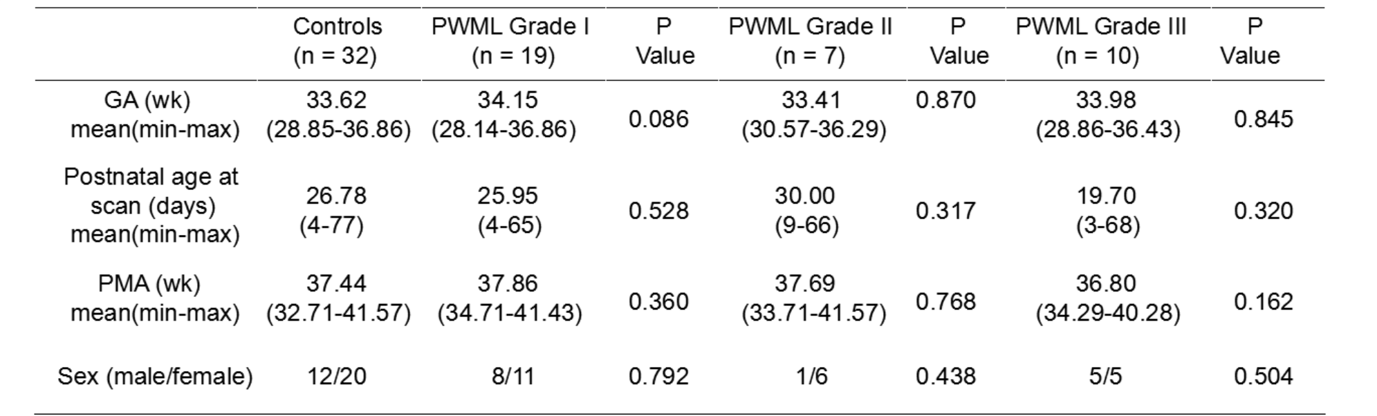
Table 1:Demographics of preterm neonates with PWMLs and controlsa. Note:—GA indicates gestational age; PMA, postmenstrual age.
a:PWML grade I,PWML grade II and PWML grade III groups are compared with the control group in GA,Postnatal age at scan,PMA and gender.
Chi-Square test was performed to evaluate the gender ratios differences across groups. Non-parametric test was used to evaluate differences in GA, postnatal age, PMA, and regional values of metrics across groups. Tests were considered significant at P<0.016 .