4660
Hemispheric asymmetry of brain motor-related white matter:a diffusion kurtosis imaging study1the First Affiliated Hospital, Xi'an Jiaotong University, Xi'an, China, 22. MR Research China, GE Healthcare, Xi'an, China, China
Synopsis
Motor function development is a necessary condition for later life. It is closely related to cognitive psychological development1. As an important white matter reflecting the motor function, clarify the asymmetry of corticospinal tract (CST) development, which is of great significance for revealing the developmental of behavior and exploring the mechanism of disease2. This study aims to use DKI parameters investigate the asymmetry of CST development. Our results suggest that from 0-13 years old, there is hemispheric asymmetry in the development of CST, and they show left-sided dominance ;the asymmetry of CST appears at 6 months.
Introduction
From birth to childhood, motor development has experienced important milestones. Such as vertical neck, sitting, standing, walking, and gradually implementing complex and fine functions3. At the same time, research have shown that sensorimotor exploration drives cognition1. The CST is of paramount importance in the motor system, as it mediates voluntary distal movements. However, the motor development has laterality, such as hand function. Therefore, as an important fiber bundle reflecting motor function, the asymmetric development of CST is of great significance to the study of basic developmental rules and disease mechanisms. Asymmetry in CST development from birth to childhood has not yet been revealed. Is it born with birth? Based on this, our research aims to explore the asymmetry of CST development based on diffusion kurtosis imaging.Methods
This study was approved by the local Internal Review Board and all parents of participants had signed the informed consents. For the inability to cooperate with the study subjects, the sedation was performed before the examination. The inclusion criteria were completed DKI examination and no significant abnormalities were observed in conventional magnetic resonance. Newborn asphyxia (5minApgar ≤ 7 points), hypoxic ischemic encephalopathy, central nervous system infection, epilepsy, developmental delay and other diseases that may affect the central nervous system are excluded.Single-shot EPI diffusion kurtosis imaging was performed on a 3.0T scanner (General Electric Signa HDXT, WI, USA) with an eight-channel head coil. The other parameters were: b values = 500, 1000, 2000, 2500 s/mm2; 18 gradient directions; Repetition time/Echo time =11000/91.7 ms; thickness = 4 mm; FOV = 180 × 180 mm2 ~ 240 × 240 mm2; acquisition matrix = 128 × 128 ~ 172 × 172. Diffusion and kurtosis tensors were estimated by using constrained weighted linear least squares. Fractional anisotropy (FA), Mean kurtosis(MK), Axonal water fraction (AWF), Tortuosity (TorT_I) were calculated according to the white matter model for DKI.
Use local template to register with JHU template to get templates and atles of different ages. Select CST_L and CST_R as regions of interests (ROI). Paired T test and non-parametric test are used to suggest CST_L and CST_R differences; The bar chart is used to show the difference between CST_L and CST_L. Tests were considered significant at P≤0.05.
Result
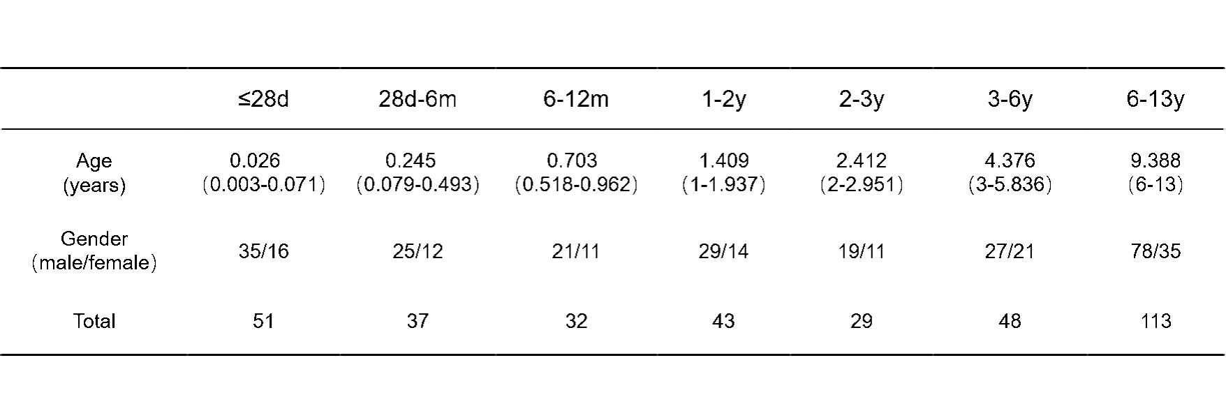
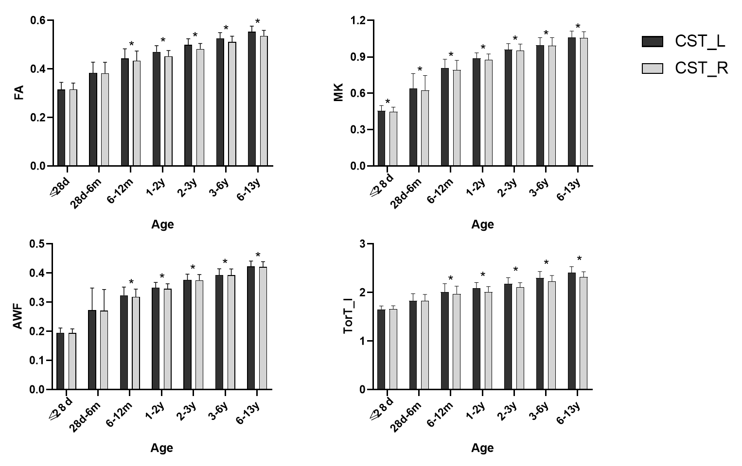
A total of 353 subjects were included. They are divided into seven groups according to the age of the examination: ≤28d,28d-6m,6-12m,1-2y,2-3y,3-6y,6-13y. (Table 1) Difference analysis between CST_L and CST_R: From 0-13 years old, the FA, MK, AWF and TorT_I of CST_L and CST_R were statistically significant, and showed left-sided dominance; But from birth to 6 months of age, only MK showed statistical difference.(Fig 1)Discussion
This study used DKI to explore the asymmetry of CST development from neonate to childhood. Different from the previous hemispheric asymmetry research, our study subjects is the continuous stage from 0-13 years old. It is found that CST have hemispheric difference and left-hemisphere advantage. Although high variability exists between individuals, the left and right hemispheres of our brain display anatomical and molecular left-right asymmetries that correlate with their functional specialization in particular cognitive processes4,5. The presence of left CST laterality after 6 months may be related to the development of motor function. For example, after 6 months of age, there are signs of hand grasping and standing.Conclusion
From 0-13 years old, there is hemispheric asymmetry in the development of CST, and they show left-sided dominance; The CST hemispheric asymmetry does not exist at birth, but appears at 6 months. From birth to 6 months of age, the difference in laterality of CST was reflected in glial cell proliferation around the axon, but there was no difference in the axon itself.Acknowledgements
This study was supported by the National Natural Science Foundation of China (81971581, 81901823, 81771810), National Key Research and Development Program of China (2016YFC0100300), the 2011 New Century Excellent Talent Support Plan of the Ministry of Education of China (NCET-11-0438), the Project Funded by China Postdoctoral Science Foundation (2019M653659), and the Natural Science Basic Research Plan in Shaanxi Province of China (2019JQ-198).References
1. Martin JH. The corticospinal system: from development to motor control[J]. Neuroscientist, 2005, 11 (2): 161-173.
2. Welniarz Q, Dusart I, Roze E. The corticospinal tract: Evolution, development, and human disorders[J]. Dev Neurobiol, 2017, 77 (7): 810-829.
3. Adolph KE, Hoch JE. Motor Development: Embodied, Embedded, Enculturated, and Enabling[J]. Annu Rev Psychol, 2019, 70: 141-164.
4. Duboc V, Dufourcq P, Blader P, et al. Asymmetry of the Brain: Development and Implications[J]. Annu Rev Genet, 2015, 49: 647-672.
5. Lee C-Y, Tabesh A, Nesland T, et al. Human brain asymmetry in microstructural connectivity demonstrated by diffusional kurtosis imaging[J]. Brain Research, 2014, 1588: 73-80.
Figures