4658
The Morphological Development of Hand Knob Areas in Children Aged 6m to 10 years1Department of Diagnostic Radiology, The First Affiliated Hospital of Xi’an Jiaotong University, Xi’an, China, 2MR Research China, GE Healthcare, Xi'an, China
Synopsis
The cortical representation of hand motor function, hand knob region, is localized in the precentral gyrus. We found that the age-related rapid development of hand knob areas peaks at 4-5 years, and the left side increases faster than right. And the different increase rate between the left and right hand knob regions of normal children may be related to dominant hemisphere of handedness. This study will help to understand the process of hand function development in children and provide the research basis for the study of the upper limb pathway of the nerve center.
Introduction
The cortical representation of hand motor function is localized in the precentral gyrus and characterized as having either an inverted omega shape or, sometimes, a horizontal epsilon shape in MR axial scans(1), which are task-state fMRI activations(2). And hand movements to the constraints of the situation in the second half of the first post-term year(3). The hand and arm function develops early and it takes many years to reach adult hand ability(3). In previous studies, several gross regions such as the precentral gyrus showed significant linear or curvilinear correlations between gray matter volume and age on an increasing trajectory(4, 5). This purpose of our study is to illustrate the age correlated trend of hand knob regions, which will help to understand the process of hand function development in children. And this research would be helpful to provide the direction for clarifying the responsible lesion of hand dysfunction.Methods
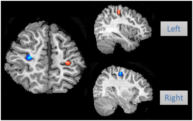
This study was approved by the local Internal Review Board and all parents of participants had signed the informed consents before MRI scanning. Patients 41 subjects (23 boys, 18 girls) with age of MR scanning during 6 months to 9.91 year-old were retrospectively evaluated without abnormalities in brain MR images. Subjects were excluded who were confirmed or suspected to have malformations of central nervous system, infections, metabolic disorders, abnormal appearances in conventional MRI and have neurologic symptom such as epilepsy. These subjects are examined by using a 3.0T scanner (Signa HDxt, GE, Milwaukee, WI, USA) with an 8-channel head coil. Data acquisition includes three-dimensional fast spoiled gradient-echo T1-weighted sequence (TR/TE, 10.42ms/4.74ms; isotropic 1×1×1mm3; thickness, 1mm) and transverse fast spin-echo T2-weighted sequence (TR/TE, 200ms/116.48ms; thickness, 4mm), and T2 Flair sequence (TR/TE, 9600ms/110.35ms; thickness, 4mm). We have hand drawn the mask of hand knob regions included each side of grey matter and white matter and saved respectively, which processed by FMRIB software library (FSL; http://www.fmrib.ox.ac.uk/fsl). (Figure.1) Then, we use SPM12 to segment and calculate the total brain volume. Finally, we respectively calculate the ratio of the gray matter volume, the white matter volume or the whole hand knob regions volume with the total brain volume. And surface area and volume MM were analyzed by software developed by the authors (AK, GE healthcare, US). The correlation analysis between age and parameters were statistically analyzed by using SPSS 18.0 (SPSS, Chicago, IL, USA); P<0.05 is considered as statistically significant difference. The inter-observer and intra-observer agreement were evaluated by using ICC analysis.Results
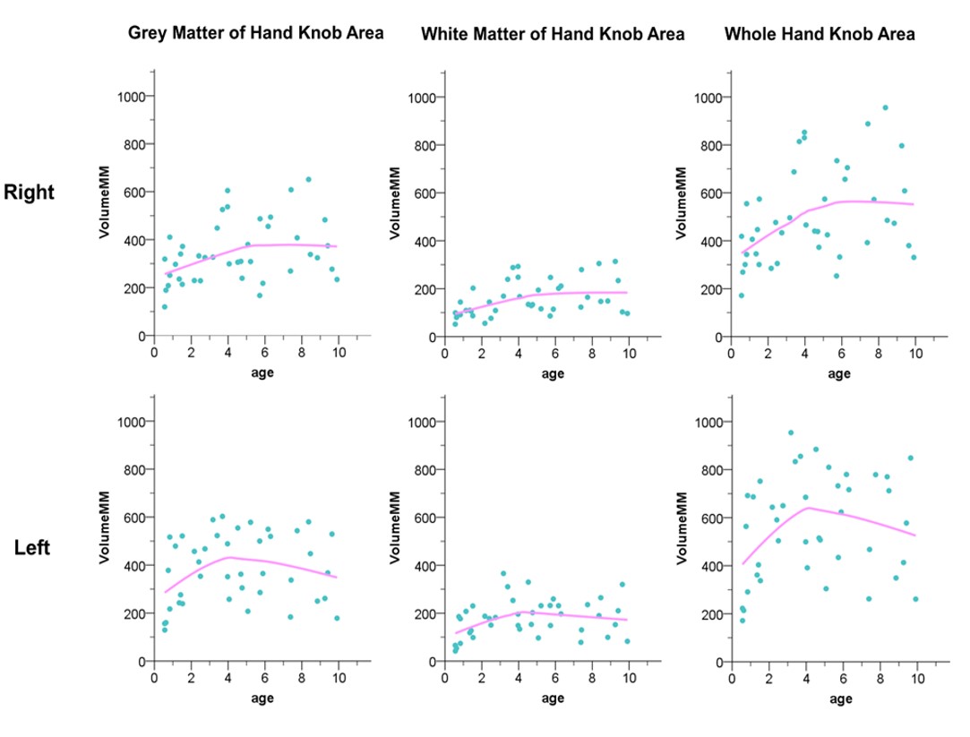
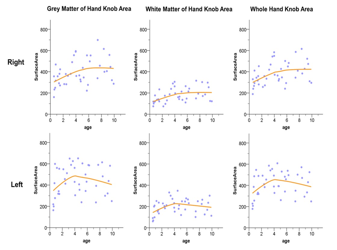
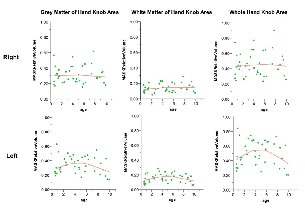
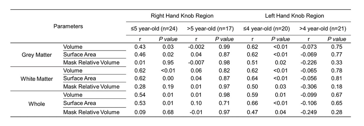
We found different age-related patterns between the bilateral hand knob areas. The volume and surface area of gray matter, white matter, and total hand knob increased with age, and the left side peak at about 4 years old while the right side peak about 5 year-olds. (Figure.2 - 4) Meanwhile, we study the correlation between the parameters of the bilateral hand knob regions and the age with the peak age as the boundary. (Table.1) Before reaching peak age, both sides of hand knob regions increased rapidly (P<0.05, r≥0.43), and the left side grew faster than the right side. Otherwise, the growth rate of the right hand knob areas was almost the same as that of the whole brain volume (P>0.05), while the growth rate of the left side was significantly faster than that of the whole brain volume before reaching the peak age (P<0.05, r≥0.47). (Figure.2 - 4, Table.1) Reproducibility between repeated measurements was high for hand knob regions (inter-observer ICC = 0.916, P<0.001; intra-observer ICC = 0.928. P<0.001).Discussion
Development processes in human brain gray matter and white matter involve different stages. Before the peak age of 4-5 years, the volume and surface of the hand knob regions are keep increasing. As the development of the hand skills, between the ages of three and six, there is a rapid improvement in fine-motor skills, such as bimanual skills, manual dexterity, eye-hand coordination, and object manipulation (6). Previous study showed cortical volume increase due to cortical thickness and surface area increasing faster before 2-year-old(6, 7). And cortical volume growth observed after 1–2 years may be driven mainly by surface area expansion. After 4-5 years, cortical thickness decreases linearly and surface area increased relatively slowly. Therefore, the cortical volume keep relatively stable after 4-5 years(7). Meanwhile, compared to gray matter, white-matter volume growth is slower and more protracted after birth (9). Otherwise, the asymmetry development process of the both sides of hand knob may be related to dominant hemisphere of handedness which is a multigenic trait seem to be established early in development(8). As fetuses move their right arms and suck their right thumbs more often than their left as early as 15 weeks, a preference that correlates with handedness later (10).Conclusion
The rapid development stage of hand knob areas is before 4-5 years which conforms to the developmental rules of fine hand movement in children. And the left hand knob region is faster and earlier than right. The age-related different processes between the left and right may be related to dominant hemisphere of handedness.Acknowledgements
This study was supported by the National Natural Science Foundation of China (81901823, 81771810, and 81971581), National Key Research and Development Program of China (2016YFC0100300), the 2011 New Century Excellent Talent Support Plan of the Ministry of Education of China (NCET-11-0438), the Project Funded by China Postdoctoral Science Foundation (2019M653659), and the Natural Science Basic Research Plan in Shaanxi Province of China (2019JQ-198).References
1.Caulo M, Briganti C, Mattei PA, Perfetti B, Ferretti A, Romani GL, et al. New morphologic variants of the hand motor cortex as seen with MR imaging in a large study population. AJNR American journal of neuroradiology. 2007;28(8):1480-5.
2.Inuggi A, Filippi M, Chieffo R, Agosta F, Rocca MA, Gonzalez-Rosa JJ, et al. Motor area localization using fMRI-constrained cortical current density reconstruction of movement-related cortical potentials, a comparison with fMRI and TMS mapping. Brain research. 2010;1308:68-78.
3.Hadders-Algra M. Early human motor development: From variation to the ability to vary and adapt. Neuroscience and biobehavioral reviews. 2018;90:411-27.
4.Taki Y, Hashizume H, Thyreau B, Sassa Y, Takeuchi H, Wu K, et al. Linear and curvilinear correlations of brain gray matter volume and density with age using voxel-based morphometry with the Akaike information criterion in 291 healthy children. Hum Brain Mapp. 2013;34(8):1857-71.
5.Li G, Lin W, Gilmore JH, Shen D. Spatial Patterns, Longitudinal Development, and Hemispheric Asymmetries of Cortical Thickness in Infants from Birth to 2 Years of Age. The Journal of neuroscience : the official journal of the Society for Neuroscience. 2015;35(24):9150-62.
6.Siu AM, Lai CY, Chiu AS, Yip CC. Development and validation of a fine-motor assessment tool for use with young children in a Chinese population. Res Dev Disabil. 2011;32(1):107-14.
7.Gilmore JH, Knickmeyer RC, Gao W. Imaging structural and functional brain development in early childhood. Nat Rev Neurosci. 2018;19(3):123-37.
8.Duboc V, Dufourcq P, Blader P, Roussigne M. Asymmetry of the Brain: Development and Implications. Annu Rev Genet. 2015;49:647-72.
9. Bullins, J., Jha, S. C., Knickmeyer, R. & Gilmore, J. in Handbook of Preschool Mental Health: Development, Disorders, and Treatment (ed. Luby, J. L.) 73–97 (The Guilford Press, New York, 2017)
10. Hepper PG, Shahidullah S, White R. 1991. Handedness in the human fetus. Neuropsychologia 29(11):1107–11
Figures