4637
The subregion scheme of the precuneus in children with autism spectrum disorder: a multimodal network analysis based on the Brainnetome Atlas1Huaxi MR Research Center (HMRRC), Department of Radiology, West China Hospital of Sichuan Uinversity, Chengdu, China, 2Psychoradiology Research Unit of the Chinese Academy of Medical Sciences (2018RU011)), West China Hospital of Sichuan Uinversity, Chengdu, China
Synopsis
To explore the brain network characteristics in children with ASD, we calculated the whole-brain structural and functional connectivity and its network topological attributes between ASD children and typical development (TD) by using whole-brain multimodal connectivity network analysis based on the Brainnetome Atlas. This study found that the left dorsomedial parieto-occipital sulcus was the area of overlap within both structural and functional connectivity abnormalities in ASD children compared with TD, implicated that this subregion of the precuneus was a key node in ASD brain network during childhood.
Introduction
Autism spectrum disorder (ASD) is a spectrum of neurodevelopmental disorders with social communication dysfunction as the core symptom. Although ASD brings a serious burden on families and society, its neuropathological mechanisms are still unclear1. Previous network researches are mostly conducted by using the Automated Anatomical Labeling (AAL) or other brain maps because its segmentation only based on general morphological markers such as sulcus and gyrus, the information about sub-regional segmentation is lacking. The Brainnetome Atlas is a brain map drawn based on living human brain imaging information, and it could provide more details about subregions information2. For example, The Brainnetome Atlas delineates four subregions in the precuneus, including dorsomedial parieto-occipital sulcus (dmPOS), ventromedial, dorsomedial and dorsolateral precuneus, which is not reported in previous brain maps like AAL. To explore the network mechanism implicated in brain subregions in ASD, we investigated the whole-brain structural and functional connectivity network based on the Brainnetome Atlas and its topological characteristics3 between ASD children and typical development (TD), by using diffusion tension imaging (DTI) and resting-state functional magnetic resonance imaging (RS-fMRI).Methods
Both of the DTI and RS-fMRI data was obtained from the dataset of New York University in Autism Brain Imaging Data Exchange (ABIDE). We first analyzed the Social Responsiveness Scale (SRS) scores in an age- and IQ-matched (range 5-12) simple of 25 ASD boys (age = 7.62 ± 1.70) and 17 TD boys (age = 8.69 ± 1.76). The imaging data was collected using a 3.0 T Simens Signa HDXT MRI scanner with an 8-channel phase array head coil, employed by a Spin echo-plannar echo imaging (SE-EPI) sequence. We conducted the preprocessing of the DTI data and constructed the white matter fiber matrix by deterministic fiber tracing by using the Diffusionkit software. And we used the BRANT (BRAinNetome Toolkit) software to preprocess the RS-fMRI data and calculated the whole-brain FC matrix based on the 273 brain regions of the Brainnetome Atlas. Then we investigated the abnormalities in functional connectivity networks and network topological properties between ASD and TD boys by using two-sample t -test, and verified the statistically significance by using Permutation Test. And we conducted correlation analysis between the network topologic attributes and SRS scores.Results
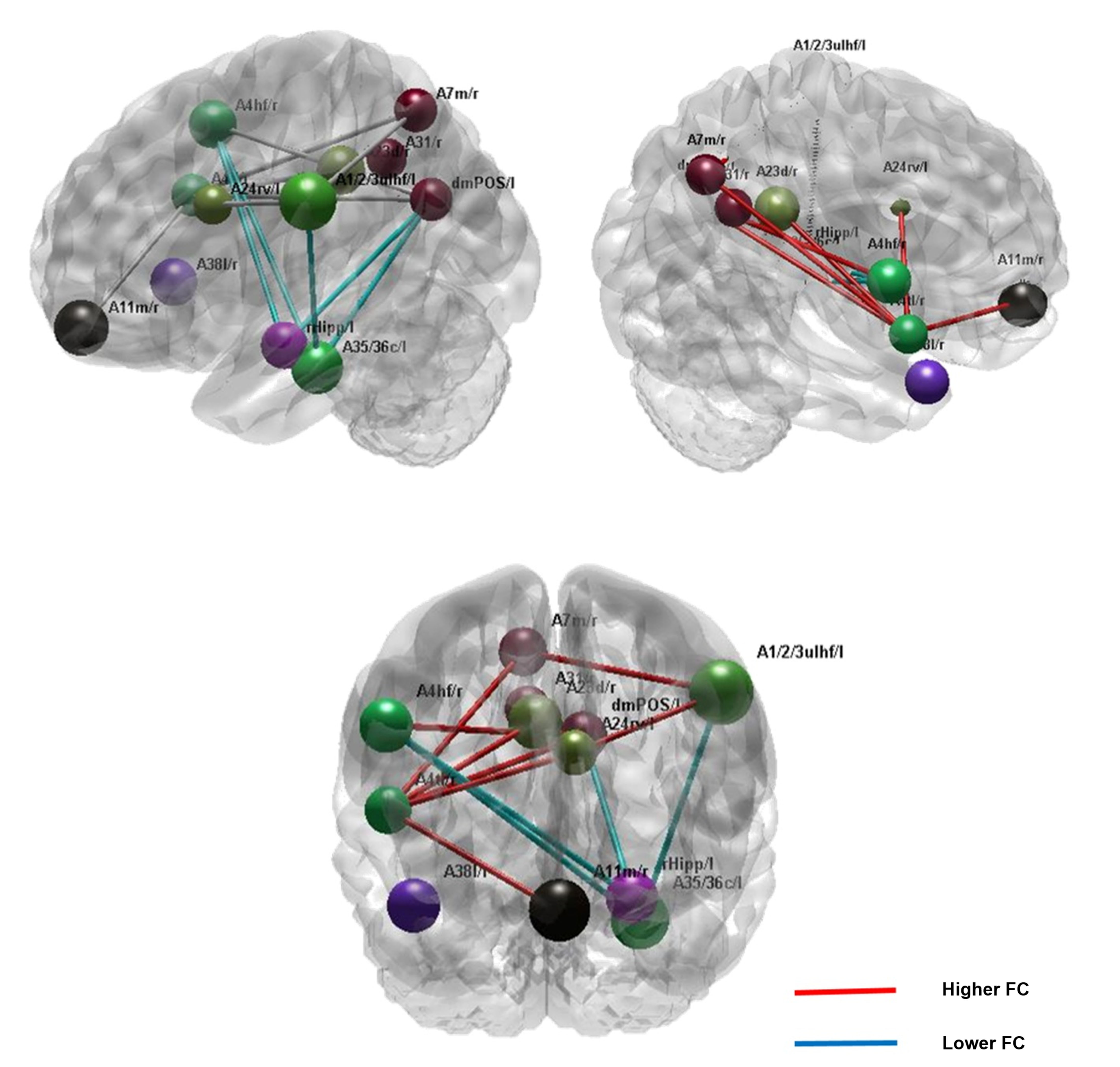
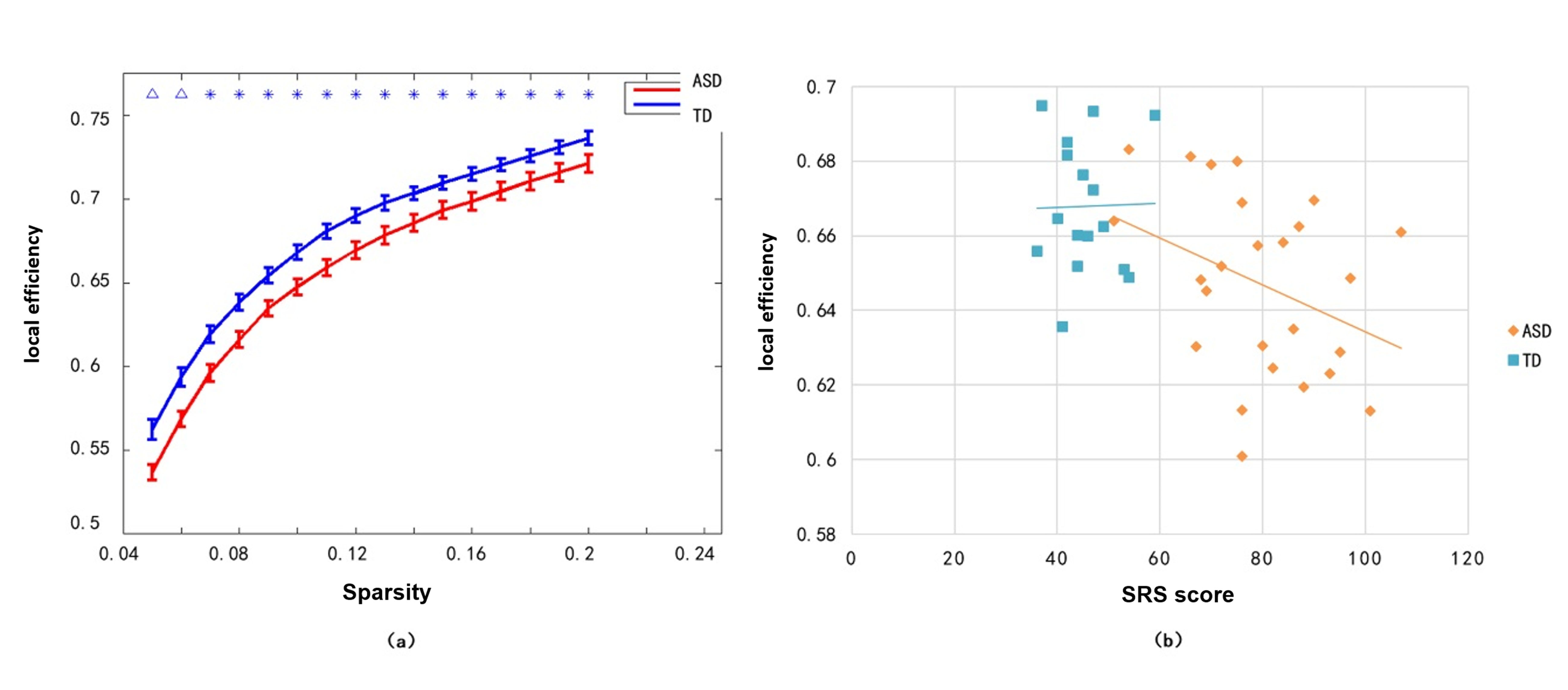
The mean SRS score in ASD boys(79.560±13.696) is statistically significantly higher than it in TD (45.375±6.163) (p = 0.007). Based on the whole-brain structural connectivity network analysis (showed in Figure 1), we found that ASD boys showed the lower whiter matter structural connectivity between the left dmPOS and inferior parietal lobule, and between the right dorsolateral precuneus and the left dmPOS than TD. And the whole-brain FC network analysis detected that the FC between the left dmPOS and hippocampus decreased in ASD boys compared with TD (as shown in Figure 2). Relative to TD, ASD boys exhibited increased FC between the left dmPOS and postcentral gyrus (upper limb and head area), and between the right ventromedial precuneus and precentral gyrus. As shown in figure 3, the local efficiency of the brain FC network in ASD boys was statistically significantly lower than that of the control group in all sparsity ranges (t∈[-2.93, -2.18]. p∈[0.001, 0.023]). And the local efficiency of the brain functional connection network was significantly negatively correlated with the total SRS score (r = -0.494 when S = 0.1).Disscussion
Our study demonstrated that the left dmPOS overlap in both structural and functional connectivity abnormalities in ASD children compared with TD, implicated that this subregion of the precuneus was a key node in ASD brain network during childhood. The precuneus is an important part of the default network which participates in self-awareness behavior4, and is activated in healthy people during the "theory of mind-related tasks."5. Our results detected the impairment of precuneus in ASD children, which was consistent with previous research that reported the brain activity in the left precuneus was reduced in 10 - to 16-year-olds with ASD when they performed the "theory of mind" task compared to a control group6. According to this multimodal connectivity network analysis, our finding demonstrated that cortical-limbic connectivity predominantly decreased while cortical-cortical connectivity mainly increased in ASD children relative to TD. Graph-based analysis of the topological properties of the brain network showed that the local efficiency of the FC network was significantly negatively correlated with the total SRS score, suggesting that the local efficiency of the FC network could be a potential marker to predict social communication ability dysfunction.Conclusion
Based on the brain network analysis by using Brainnetome Atlas, we found that the left dmPOS, as a sub-region of precuneus, which was a high weight node in ASD children, suggesting that sub-regions of precuneus played separated roles in the ASD children. In summary, we systematically explored the features of both brain structural and functional connectivity networks in ASD children compared with HC by using whole-brain connectivity network analysis based on Brainnetome Atlas, providing new evidence for exploring the neural network mechanism of autism.Acknowledgements
The authors declare that they have no conflicts of interest. This study was supported by the Psychoradiology Research Unit of the Chinese Academy of Medical Sciences.Dr. Gong would also like to acknowledge the support from his Changjiang Scholar ProfessorshipAward (Award No. T2014190)References
1. Lai MC, Lombardo MV, Baron-Cohen S. Autism. Lancet (London, England) 2014; 383: 896-910.
2. Fan L, Li H, Zhuo J, et al. The Human Brainnetome Atlas: A New Brain Atlas Based on Connectional Architecture. Cerebral Cortex 2016; 26: 3508-26.
3. Yap P-T, Wu G, Shen D. Human Brain Connectomics: Networks, Techniques, and Applications. Ieee Signal Processing Magazine 2010; 27: 131-4.
4. Abbott AE, Nair A, Keown CL, et al. Patterns of Atypical Functional Connectivity and Behavioral Links in Autism Differ Between Default, Salience, and Executive Networks. Cerebral Cortex 2016; 26: 4034-45.
5. Bagozzi RP, Verbeke WJMI, Dietvorst RC, Belschak FD, van den Berg WE, Rietdijk WJR. Theory of Mind and Empathic Explanations of Machiavellianism: A Neuroscience Perspective. Journal of Management 2013; 39: 1760-98.6.
6. Kana RK, Maximo JO, Williams DL, et al. Aberrant functioning of the theory-of-mind network in children and adolescents with autism. Molecular Autism 2015; 6.7.
Figures
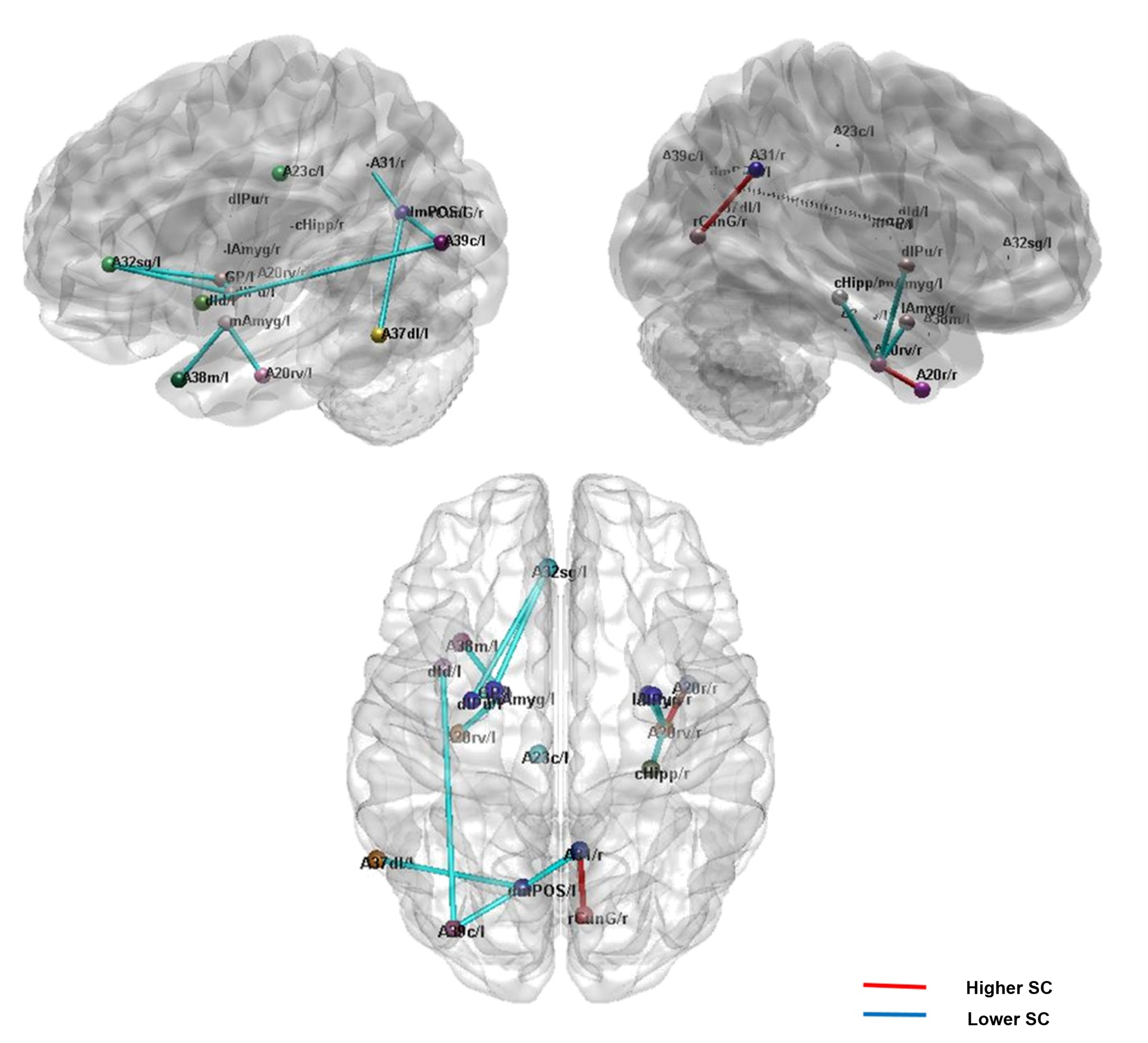
Figure 1. Brain white matter structural connectivity alteration in ASD children compared with TD based on the Brainnetome Atlas. Red: higher SC; blue: lower SC; A31: dorsolateral precuneus; A32sg: subgenual anterior cingulate cortex;A37dl: dorsolateral middle temporal gyrus;A39c: caudate inferior parietal lobule; ASD: Autism spectrum disorder; dmPoS: dorsomedial parieto-occipital sulcus;l:left;lAmyg: lateral amygdala;r:right,;rCunG: rostral cuneus; SC: structural connectivity; TD: typical development.