4631
Dorsal-ventral stream development for visual word processing1Department of ENT, Escorts Heart Institute and Research Center, New Delhi, India, 2Former Department of NMR and MRI Facility, Former ALL INSTITUTE OF MEDICAL SCIENCES, NEW DELHI, India, 3Department of NMR and MRI Facility, All India Institute of Medical Sciences, New Delhi, India, 4Department of Psychiatry, All India Institute of Medical Sciences, New Delhi, India, 5Department of Neurology, Fortis Hospital, New Delhi, India, 6Department of Psychiatry (Psychology Unit), All India Institute of Medical Sciences, New Delhi, India, 7Department of Linguistics, School of Language, Jawahar Lal Nehru University, New Delhi, India, 8Department of Biostatistics, All India Institute of Medical Sciences, New Delhi, India
Synopsis
Reading skill is window to the world that we acquire with development. Dual-route model predicts our proficiency of grapheme (text) to phoneme (sound) conversion and semantic decoding (understanding content) for visual words. Fast processing by frontoparietal pathway while meaningful word reading but lexical decision to pseudowords proceeds slowly and exhaustively. Pseudoword successful orthographic mapping recruits ventral and then dorsal areas. This phonological proficiency and exhaustive mental lexicon search during reading is automatized with skill development. Current study evidences recruitment of inferior frontal gyrus (BA44 and 45), insula, thalamus, caudate nucleus and cortical reorganizations of skill developmental with age
Introduction
Learning to read requires phonological interface with lexicosemantic and orthographic features [1, 2]. Computation of orthographic input (visual word) [3] is facilitated by appropriate lexical selection from semantic mental storage [4]. Dual--route model explains fast mapping visually (orthographic representations), the words from pseudowords onto stored word-form representations [1, 3] Early readers rely on phonology-based grapheme-phoneme conversion attributed to left dorsal temporoparietal circuit [5]. Efficient readers involve automatic visual-orthographic lexical decoding due to communication between frontal and posterior areas [5, 6, 7]. Coordinated interplay of the dorsal-ventral streams during development is proportionate with reading efficiency [6, 7]. So the present study explored the dual-route model during development by comparing the word-pseudoword processing of typical reading children and adults.Methods
The study was carried out on healthy typical reading children (CH; n= 20; age range 8 to 15 years) and adult subjects (AD; n=16; age range 25 to 45 years) after IEC approval. Inclusion criteria were: right handedness, high proficiency in reading Hindi (shallow orthography) and given written consent. Exclusion criteria were left handedness, any sensory impairment (hearing/ vision), neurological or psychiatric problems, and any contraindication for MRI. The Blood oxygen level dependent (BOLD) data was acquired with clinical 3T whole body MR scanner with 32 channel head coil (Achieva 3.0T TX, Philips, Netherlands). Single-shot echo planar imaging (EPI) sequence was used with slice thickness 5 mm, number of slices = 29, TR: 2000 ms, TE: 30 ms, flip angle = 90°, FOV = 230 mm, Dynamics: 222, Resolution: 64x64, overlaid on render template. The visual text stimuli were presented using Eprime (version 1.1, Psychology Software Tools Inc, USA) and MR compatible LCD monitor (NordicNeuroLab, Norway). The task comprised of Meaningful words (MW) (5 event x 4 blocks) and Pseudowords (PW)(5x4) in Hindi where each event was of 2.5 sec duration. The paradigm included baseline 28 sec (i.e. background noise with black screen display), then block of MW of 2-3 syllables, second baseline 28 sec followed by block of PW (2-3), similarly ABCABC four cycles. The response was oral reading of the text displayed and the total duration was 444 sec. Pre- and post-processing was done using SPM8 (Wellcome Department of Cognitive Neurology, London, UK). The BOLD clusters were converted from MNI template to the Talairach and Tornoux coordinates, for estimation of anatomical areas. The group data was analyzed by one-way ANOVA test (p<0.001, cluster threshold 10).Results and Discussion
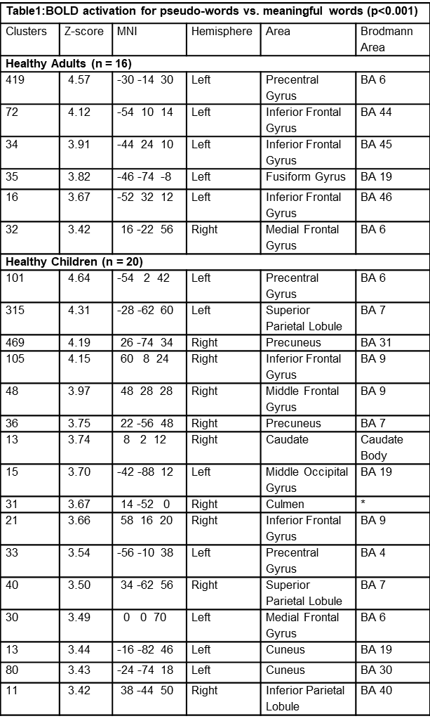
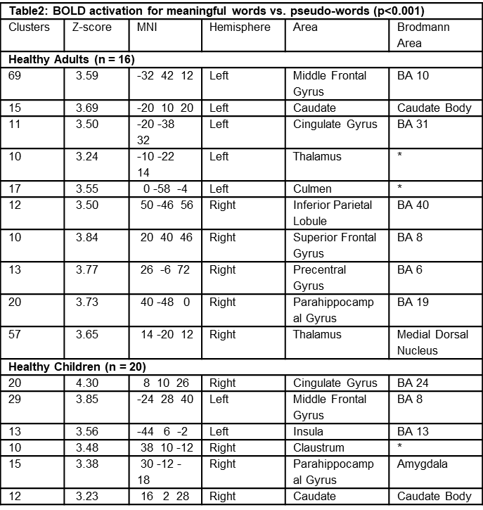
Hindi is orthographically shallow language where readers rely on phonology-based processes (i.e., grapheme-phoneme conversion) unlike English (deep orthography) its whole-word recognition (may have ambiguous grapheme-phoneme mapping [5]. The BOLD data in adult (AD) participants showed that on comparison of MW vs PW left hemispheric dominance of middle frontal gyrus (MFG), caudate, cingulated gyrus, culmen and right dominance of inferior parietal, superior frontal, precentral gyrus. On comparison of PW vs MW left precentral, inferior frontal gyrus (BA44, BA 45 and BA46), fusiform and right medial frontal gyrus showed significant BOLD activity. The fusiform gyrus provides access code to semantic or phonological information represented in posterior left middle temporal gyrus (MTG) [5. 6]. Neural activity in the left inferior frontal gyrus (IFG, BA44), anterior insula, thalamus and caudate nucleus, supports grapheme to phoneme conversion during visual word processing [2, 5]. BOLD activity for PW vs MW in healthy children (CH) was similar to adults but was represented bilaterally (Table 1 and Table 2) [8]. Group comparison (Children vs Adults analysis) MW show anterior cingulate and PW show insula activation. Dorsal-ventral participation for meaningful words and dorsal control for pseudowords differences in adults and children is due to non-automatized (Figure 1) [5]. This study suggests that differences in dorsal pathway activation for PW in two groups are due to non-atomization of reading skill in children [5, 9].Conclusion
The results indicate that pseudoword reading reflects the need for a more exhaustive search in the mental lexicon. The study evidences the dual-route model of lexical decision and there is switching of faster pathway for grapheme to phoneme conversion. Automatization of reading skill during development with age is attributed to neural reorganizations.Acknowledgements
No acknowledgement found.References
[1] Krishnamurthy LC, Krishnamurthy V, Crosson B, Rothman DL, Schwam DM, Greenberg D, Pugh KR, Morris RD. Strength of resting state functional connectivity and local GABA concentrations predict oral reading of real and pseudo-words. Sci Rep. 2019 Aug 6;9(1):11385. doi: 10.1038/s41598-019-47889-9.
[2] Price CJ. A review and synthesis of the first 20 years of PET and fMRI studies of heard speech, spoken language and reading. Neuroimage. 2012; 62(2):816-47.
[3] Bub D. N., and Arguin M., 1995. Visual word activation in pure alexia. Brain and Language, 49, 77- 103.
[4]Fiebach C.J., Friederici A.D., Mu¨ller K., and Yves von Cramon D., 2002. fMRI evidence for dual routes to the mental lexicon in visual word recognition. J of Cogni NeuroSc 14:1,11- 23
[5] Martin A, Schurz M, Kronbichler M, Richlan F. Reading in the brain of children and adults: a meta-analysis of 40 functional magnetic resonance imaging studies. Hum Brain Mapp. 2015;36(5):1963-81.
[6] Turkeltaub PE, Gareau L, Flowers DL, Zeffiro TA, Eden GF. Development of neural mechanisms for reading. Nat Neurosci. 2003;6(7):767-73.
[7] Wise-Younger J, Tucker-Drob E, Booth JR. Longitudinal changes in reading network connectivity related to skill improvement. Neuroimage. 2017;158:90-98.
[8] Liebig J, Froehlich E, Morawetz C, Braun M, Jacobs AM, Heekeren HR, Ziegler JC. Neurofunctionally dissecting the reading system in children. Dev Cogn Neurosci. 2017;27:45-57.
[9] Levy J, Pernet C, Treserras S, Boulanouar K, Aubry F, Démonet JF, Celsis P. Testing for the dual-route cascade reading model in the brain: an fMRI effective connectivity account of an efficient reading style. PLoS One. 2009;4(8):e6675.
Figures
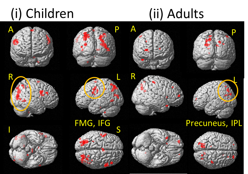
BOLD (fMRI) activation during visual word processing in healthy adults, typical reading (healthy children)
BOLD activation (important areas marked as yellow circles) rendered on standard template for visual processing (searching missing part in the picture) using one-way ANOVA (p < 0.001, voxel threshold: 10, 1 voxel: 2x2x2 mm3) in (i) healthy adults (n = 16), (ii) healthy age-matched (healthy controls, n = 20) {A- anterior view; P- posterior; R-right; L- left; I- inferior; S- superior view; MFG-middle frontal gyrus, IFG-inferior frontal gyrus, IPL- inferior parietal lobule}