4618
8-channel dipole array for 7 Tesla neonatal brain and cardiac MRI1Biomedical Engineering Department, School of Biomedical Engineering and Imaging Sciences, King's College London, London, United Kingdom, 2MR Research Collaborations, Siemens Healthcare Limited, Frimley, United Kingdom, 3Centre for the Developing Brain, School of Biomedical Engineering and Imaging Sciences, King's College London, London, United Kingdom, 4Department of Radiology, Leiden University Medical Center, Leiden, Netherlands
Synopsis
An 8-channel dipole array was investigated for neonatal brain and cardiac MR imaging at 7T. The coil array was compared using electromagnetic field simulations to a single-channel birdcage coil on a realistic baby model for brain and cardiac imaging. B1+-efficiency and SAR10g were evaluated. The dipole coil array demonstrated significant lower SAR10g levels for both imaging positions. The working of the constructed dipole array was assessed on adult head. We conclude that dipole arrays are a valuable approach for neonatal brain and cardiac MR imaging at 7T, and could be part of future ethics approval of a neonatal MRI application.
Introduction
Neonatal MR imaging at 7T could benefit from the higher achievable resolution [1] and increased SNR [2] compared to lower field MR scanners. However, there are currently no dedicated RF transmit coils for neonatal brain and cardiac MR imaging. Dipoles were demonstrated to perform efficiently for adult brain imaging in terms of B1+-field and coverage [3,4]. In this study, an 8-channel parallel-transmit dipole array was modeled and built for neonatal brain and cardiac MR imaging at 7T. The coil array was compared in simulations with a standard birdcage coil in terms of B1+ field efficiency and SAR10g levels.Methods
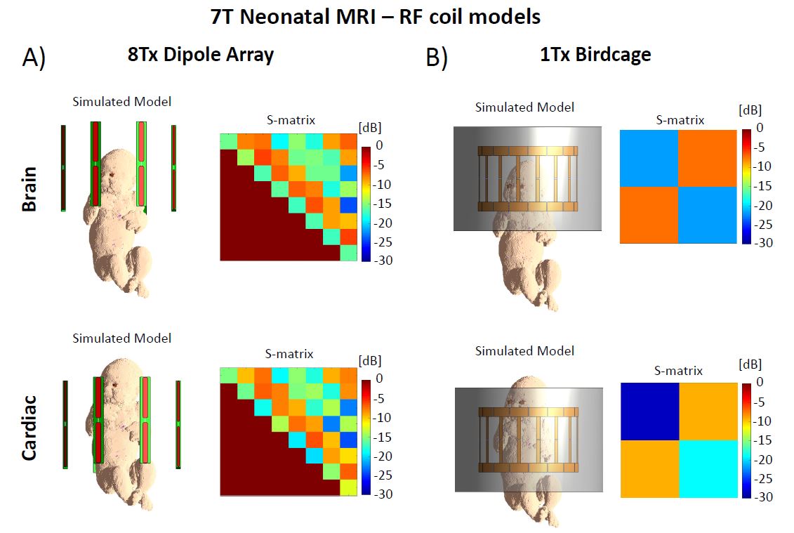
An in-house designed 8-channel dipole array was simulated for scattering-matrices (S-matrix), B1+ maps, maximum local SAR value and transmit efficiency ($$$B_1^+/\sqrt{SAR_{10g,max}}$$$) on babies for brain and cardiac MRI, and compared with a single-channel band-pass birdcage RF coil (16-legs, diameter:270mm,length:175mm) driven in quadrature using finite-difference time-domain (FDTD) simulation software (Sim4Life 5.0,ZMT,Switzerland). The birdcage design was based on commercially available adult head birdcage coil (Nova Medicals,Inc.). Both coils were tuned to 297.2MHz and matched to 50Ohms for the baby’s brain, using a co-simulation approach (Optenni Ltd,Finland) and an in-house developed baby model including 13 tissues with their corresponding dielectric properties. The computed matching parameters (circuits and component values) were then used for both brain and cardiac imaging positions (Fig.1). RF phases were optimized for the 8Tx dipole array over the brain and the heart, using a particle-swarm optimization method [5]. All measured and simulated B1+, SAR and transmit efficiency maps were normalized to 1W total input power for comparison, using the shimmed phases when evaluating the 8Tx dipole array.The in-house constructed 8-channel transmit/receive dipole array consisted of eight center-shortened dipole antennas (length:230mm, width:15mm) with each dipole etched from 35 μm copper on a FR-4 substrate (thickness:1.6mm) (Fig.4B-C). An in-house 3D-designed (Solidworks,Dassault Systemes) coil holder (inner diameter:286mm,length:390mm) was printed in polycarbonate and painted with an insulating lacquer meeting the international standards in terms of toxicity (Deed3D Technology Co., China). The coil holder was designed to be separated in two halves in order to easily position the baby for a MRI scan. Dipoles were tuned and matched (S11>-15dB) using fixed-value capacitors (American Technical Ceramics, USA) and hand-wounded copper-wire inductors.
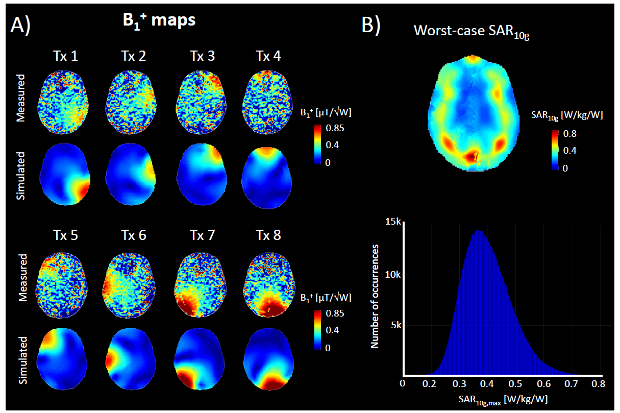
Individual B1+ maps from a healthy adult volunteer were acquired on a 7T MR scanner with 8x2kW RF-amplifiers (MAGNETOM Terra,Siemens,Erlangen,Germany) in prototype research configuration, using a pre-saturation turbo-flash sequence [6] , and compared to the simulated results using the in-house built 8-channel dipole array. SAR matrices were computed for 10g tissue mass-average and worst-case SAR10g [7] was calculated for adult head [8]. Virtual observations points (VOPs) were derived from the SAR matrices calculation, and maximal SAR10g values were computed for 1 million random RF shim sets [9].
Results
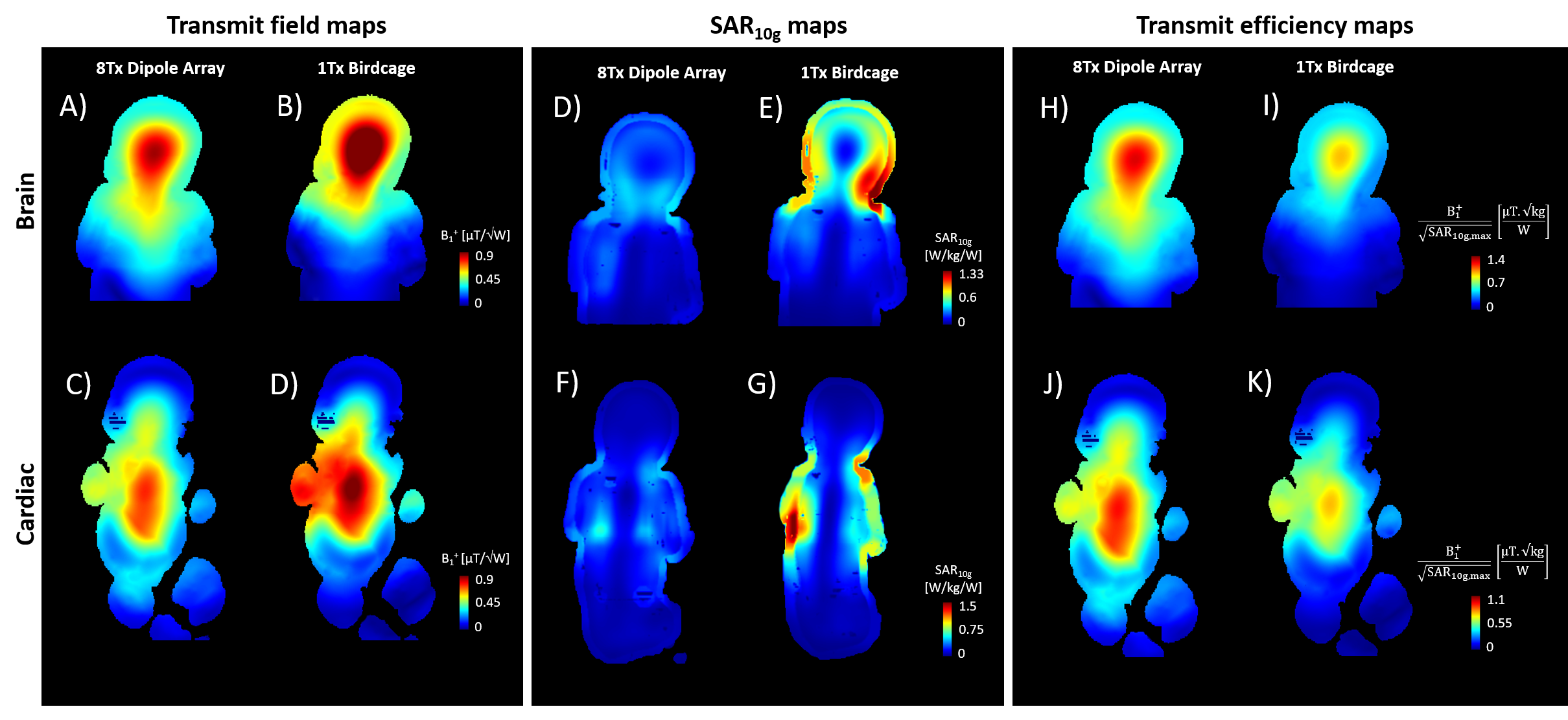
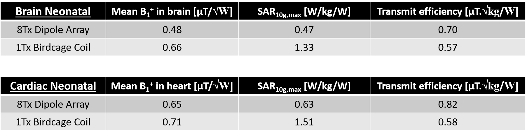
The scattering matrix for the 8Tx dipole array was similar in both brain and heart positions, while for the birdcage small variations in coupling and reflection coefficients were observed (Fig.1). The average transmit B1+ fields values achieved for the 8Tx dipole array were 27% and 8% lower than the birdcage coil for brain (Fig.2A-B) and cardiac (Fig.2C-D) positions, respectively (Table1). Nevertheless, in both brain and cardiac configurations the 8Tx dipole array demonstrated a slightly larger longitudinal coverage of the neonatal body (Fig.2A&C).For neonatal brain, the maximal SAR10g value was found in the neck region for each coil (Fig.2D-E). However, the peak SAR10g value for the 8Tx dipole array was 65% lower compared to the birdcage coil (Fig.2D-E,Table1). For neonatal cardiac MRI, the maximal SAR10g value was increased by 34% for the 8Tx dipole array while it was increased by 14% for the birdcage (Fig.2F-G,Table1) compared to the brain position.
In terms of transmit efficiency ($$$B_1^+/\sqrt{SAR_{10g,max}}$$$), the 8Tx dipole array demonstrated a 23% gain compared to the birdcage coil for brain imaging (Fig.2H-I,Table1). The difference was larger for cardiac imaging, where the 8Tx dipole array outperformed the 1Tx birdcage coil by 41% in terms of transmit efficiency (Fig.2J-K,Table1).
Measured and simulated individual B1+-field distributions were comparable in adult brain (Fig.5A). The worst-case local SAR10g, normalized to 1W total input power, was found to be around 0.8W/kg/W, and the distribution over 1 million RF shims demonstrated that 95% of the computed SAR10g,max values are actually below 0.55W/kg/W (Fig.5B).
Discussions and Conclusion
In this study, an 8-channel dipole coil array was evaluated in simulation for neonatal brain and cardiac MR imaging at 7T. Compared to a standard single-channel birdcage coil, the 8Tx dipole array demonstrated lower averaged B1+-field values in both brain and cardiac positions. Nevertheless, it outperformed the birdcage coil in terms of transmit efficiency as the maximum local SAR10g for RF shimmed phases was significantly lower. Moreover, the RF phase shimming optimization did not include SAR minimization which could potentially further decrease the power deposition in baby.The working of the in-house built 8Tx dipole array was then verified on adult brain through the correlation between simulated and measured B1+ maps for the worst-case local SAR conditions.
We conclude that dipole arrays are a valuable RF coil design approach for neonatal MR imaging of brain or heart at 7T, and could be included for the future ethics approval of a neonatal MRI application.
Acknowledgements
This work was supported by KCL 2018-2019 Partnership Fund, the Wellcome EPSRC Centre for Medical Engineering at Kings College London (WT 203148/Z/16/Z) and by the National Institute for Health Research (NIHR) Biomedical Research Centre based at Guy’s and St Thomas’ NHS Foundation Trust and King’s College London. The views expressed are those of the authors and not necessarily those of the NHS, the NIHR or the Department of Health.References
[1] Webb et al, Int. J. Imaging
Systems and Technology 20(1), 2010
[2] Vaughan et al, Magn. Res. Med. 46(1), 2001
[3] Clement et al, Magn. Res. Med. 81(2), 2019
[4] Clement et al, Magn. Res. Med. 82(3). 2019
[5] Clément et al, ESMRMB, Abstract 503, 2015
[6] Fautz et al. ISMRM, Abstract 1247, 2008
[7] Ipek O. et al, Magn. Res. Med. 71(4), 2013
[8] Gosselin M. et al, Phys. Med. Biol. 59(18),
2014
[9] Eichfelder G. et al, Magn.
Res. Med. 66(5), 2011
Figures