4599
Depicting White Matter Developmental Trajectory Using Diffusional Kurtosis Imaging:from neonate through childhood1the First Affiliated Hospital, Xi'an Jiaotong University, Xi'an, China, 22. MR Research China, GE Healthcare, Xi'an, China, China
Synopsis
Brain development is a complex process linked with behavioral, emotional, cognitive, especially the maturation process of WM is both of great scientific and clinical importance1. However, the lack of continuous development of WM from neonate to childhood. Diffusion kurtosis imaging(DKI)is a diffuse imaging method that reflects non-Gaussian distribution information of biological water. This study aimed to use DKI parameters depicting developmental trajectory of WM. The fractional anisotropy(FA), mean kurtosis(MK),axial kurtosis(AK),radial kurtosis(RK)positively correlated with age; the axial diffusivity(AD) radial diffusivity(RD) negativly correlated with age. These parameters changed rapidly before the age of 2 years old, and then gradually slowed down.
Introduction
From the newborn to childhood, a variety of lesions involving WM can occur2. However, how to judge the WM damage? Due to the differences between China and the West, we need normal WM developmental trajectory as a reference. Unlike other methods, DKI is non-Gaussian and its information is closer to biological characteristics3. We can get the following parameters from this model: fractional anisotropy(FA), mean kurtosis(MK),axial kurtosis(AK), radial kurtosis(RK), axial diffusivity(AD), radial diffusivity(RD). The goal of this study was to observe the correlation of these parameters with age, to depict WM developmental trajectory.Methods
This study was approved by the local Internal Review Board and all parents of participants had signed the informed consents. For the inability to cooperate with the study subjects, the sedation was performed before the examination. The inclusion criteria were completed DKI examination and no significant abnormalities were observed in conventional magnetic resonance. Newborn asphyxia (5minApgar ≤ 7 points), hypoxic ischemic encephalopathy, central nervous system infection, epilepsy, developmental delay and other diseases that may affect the central nervous system are excluded.Single-shot EPI diffusion kurtosis imaging was performed on a 3.0T scanner (General Electric Signa HDXT, WI, USA) with an eight-channel head coil. The other parameters were: b values = 500, 1000, 2000, 2500 s/mm2; 18 gradient directions; Repetition time/Echo time =11000/91.7 ms; thickness = 4 mm; FOV = 180 × 180 mm2 ~ 240 × 240 mm2; acquisition matrix = 128 × 128 ~ 172 × 172. Diffusion and kurtosis tensors were estimated by using constrained weighted linear least squares. Fractional anisotropy (FA), mean kurtosis(MK),axial kurtosis(AK) ,radial kurtosis(RK) ,axial diffusivity (AD), radial diffusivity (RD)were calculated according to the white matter model for DKI.
Use local template to register with JHU template to get templates and atles of different ages. Ten regions of interests (ROI) were selected including projection fibers, combined fibers, and tie fibers: left anterior thalamic radiation (ATR_L), right anterior thalamic radiation(ATR_R), left corticospinal tract (CST_L), right corticospinal tract (CST_R), genu of corpus callosum(GCC),splenium of corpus callosum(SCC), left inferior longitudinal fasciculus(ILF_L),right inferior longitudinal fasciculus(ILF_R),left superior longitudinal fasciculus(SLF_L),right superior longitudinal fasciculus(SLF_R). Using MATLAB to extract different parameter values of ROI. Spearman correlation was used to assess the correlation of parameter values with age; LOWESS was used to depict WM developmental trajectory. Tests were considered significant at P≤0.05.
Result
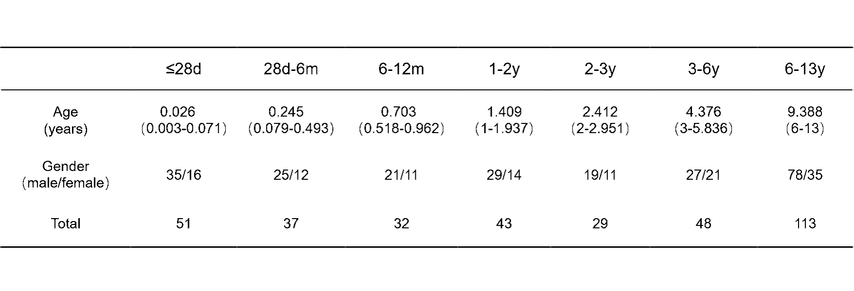
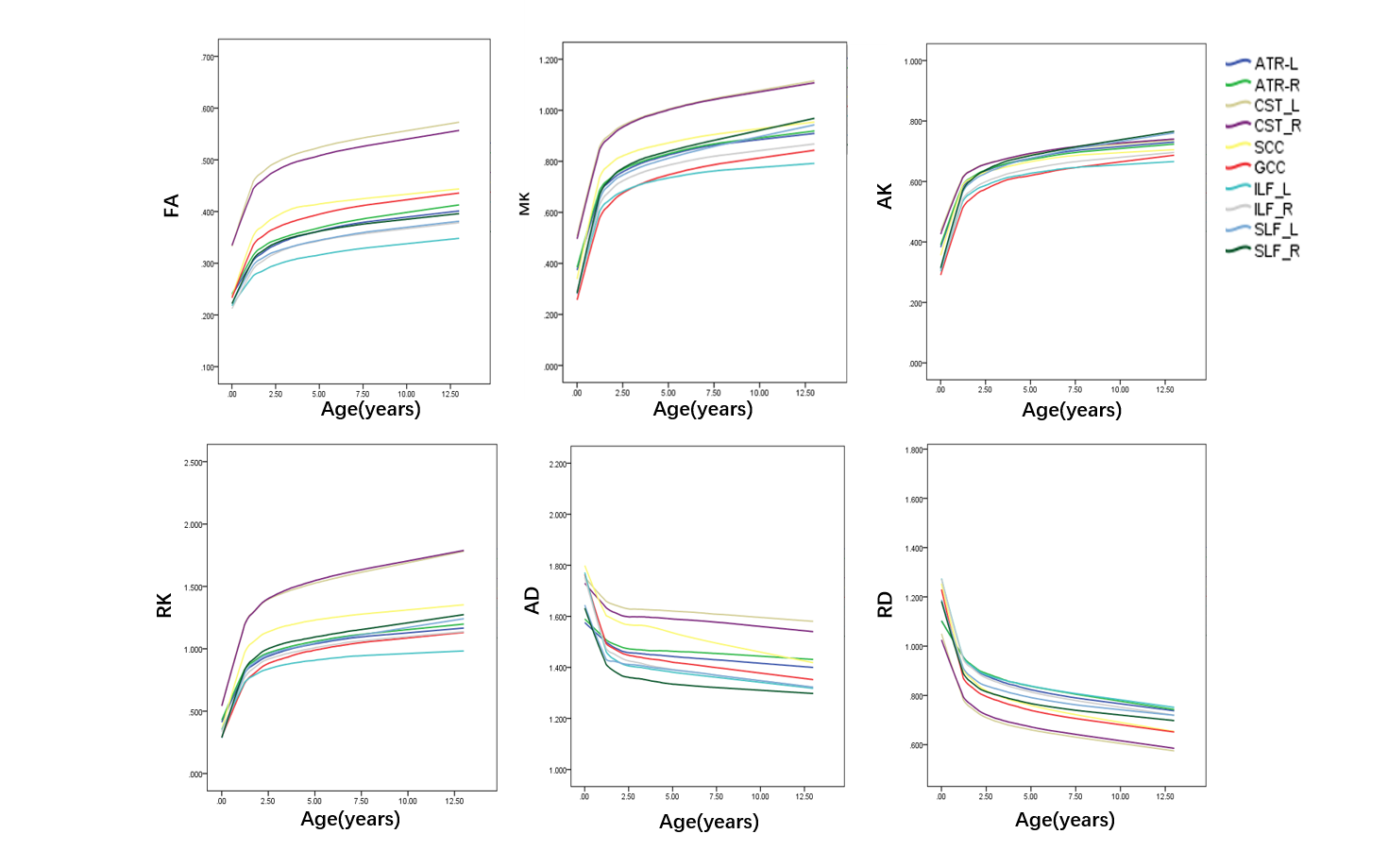
A total of 353 subjects infants were included. They are divided into seven groups according to the age of the examination:<28d,28d-6m,6-12m,1-2y,2-3y,3-6y,6-13y(Table 1).Correlation analysis:FA, MK, AK, RK were strongly correlated with age;AD and RD were negatively correlated with age (Table 2). The WM developmental trajectory shows that FA, MK, AK and RK of each ROI increase with age, AD and RD decrease with age; compared with FA, MK increases more obviously, compared with AD, RD decrease more obvious. At the same time, we observed that the values of the parameters changed rapidly before the 2 years old, and then slowly changed (Fig 1).
Discussion
Different from the previous white matter development research, our study subjects is the continuous stage from neonate to childhood. The change trend of WM development is observed by DKI parameters3,4,5. Rapid development before the 2 years old is consistent with previous results6. It suggest that the basic structural and functional framework of the human brain is in place by the second year of life, after age 2 years is characterized mainly by reorganization, ‘fine-tuning’, plasticity and remodeling of the major circuits and networks that are already established5.Conclusion
Increase with age, myelination gradually matures, and 2 years old is a developmental inflection point.Acknowledgements
This study was supported by the National Natural Science Foundation of China (81971581, 81901823, 81771810), National Key Research and Development Program of China (2016YFC0100300), the 2011 New Century Excellent Talent Support Plan of the Ministry of Education of China (NCET-11-0438), the Project Funded by China Postdoctoral Science Foundation (2019M653659), and the Natural Science Basic Research Plan in Shaanxi Province of China (2019JQ-198).References
1. Lebel C, Walker L, Leemans A, et al. Microstructural maturation of the human brain from childhood to adulthood[J]. Neuroimage, 2008, 40 (3): 1044-1055.
2. Wu EX, Cheung MM,et al. MR diffusion kurtosis imaging for neural tissue characterization[J]. NMR in Biomedicine, 2010, 23 (7): 836-848.
3. Paydar A, Fieremans E, Nwankwo JI, et al. Diffusional Kurtosis Imaging of the Developing Brain[J]. American Journal of Neuroradiology, 2014, 35 (4): 808-814.
4. Grinberg F, Maximov, II, Farrher E, et al. Diffusion kurtosis metrics as biomarkers of microstructural development: A comparative study of a group of children and a group of adults[J]. Neuroimage, 2017, 144 (Pt A): 12-22.
5. Collier Q, Veraart J, Jeurissen B, et al. Diffusion kurtosis imaging with free water elimination: A bayesian estimation approach[J]. Magnetic Resonance in Medicine, 2018, 80 (2): 802-813.
6. Gilmore JH, Knickmeyer RC, Gao W. Imaging structural and functional brain development in early childhood[J]. Nat Rev Neurosci, 2018, 19 (3): 123-137.
Figures

AD,RD:×10−3mm2/s
Fig 1.White Matter Developmental Trajectory: FA, MK, AK and RK of each ROI increase with age, AD and RD decrease with age; compared with FA, MK increases more obviously, compared with AD, RD decrease more obvious.