4596
Investigating the change of optic radiation in Septo-optic dysplasia patients with DTI
Xiaoyu Wang1, Cong Tian1, Xinxin Tao1, Mengxuan Li1, Yuli Zhang1, Xiaocheng Wei2, and Jian Yang3
1The first affliated hospital of Xi'an Jiaotong University, Xi'an, China, 2MR Research China, GE Healthcare, Beijing, China, 3Radiology, The first affliated hospital of Xi'an Jiaotong University, Xi'an, China
1The first affliated hospital of Xi'an Jiaotong University, Xi'an, China, 2MR Research China, GE Healthcare, Beijing, China, 3Radiology, The first affliated hospital of Xi'an Jiaotong University, Xi'an, China
Synopsis
Septo-optic dysplasia (SOD) is a rare congenital malformation disorder and have equally prevalent in males and females in early brain development. This study evaluated the micro-structure of optic radiation pathway using automatic fiber bundle quantization tracking (AFQ)in DT image to investigate the possible developmental impairment of optic radiation pathway that might cause by the abnormalanatomy structure of SOD patient. Result showed abnormal changes of optic radiation were found in SOD patients. The DTI tractography result could provides a non-invasive mapping of the optic radiation pathway and locate the injury.
Introduction
Septo-optic dysplasia (SOD), also known as de Morsier syndrome,1is a rare congenital malformation disorder and has a reported incidence of 1/10,1000 and is equally prevalent in males and females in early brain development.3 The symptoms are traditionally defined by three characteristic features: underdevelopment of the optic nerve, pituitary gland dysfunction, and absence of the septum pellucidum. A minimum of two features above need to be presented for clinical diagnosis.2 Previous studies have been reported on MRI structures abnormality, clinical features and possible genetic changes that could cause SOD.4-7 However, very little study have been found on the association between SOD and microstructure changes of white matter in children, in particular, the optic radiation, a key white matter structure conveying visual information from the lateral geniculate nucleus (LGN) to the primary visual cortex in the occipital lobe.8 The aim of this study was to explore the association between optic radiation structure and SOD and weather this pathway structure can be effected by SOD using the diffusion tensor imaging (DTI) tractography, which allows non-invasive mapping of the optic radiation pathway.Method:
The institutional review board of the First Affiliated Hospital of Xi’an Jiaotong University approved this prospective study, and the written informed consent were obtained from all participants. SubjectsThis study enrolled 4 patients with SOD (aged 3-5, 3 males) and 4 healthy control (aged 3-5, 3 males) who performed MRI at the Department of Diagnostic Radiology in our hospital between January 2016 to October 2018. Data on visual acuity, nystagmus and imaging feature such as optic nerve hypoplasia and absent of septum pellucidum were collected. MR ProtocolsAll subjects were examined by using a 3.0T scanner (Signa HDxt, General Electric Medical System, Milwaukee, WI, USA) with an 8-channel head coil. Data acquisition included three-dimensional fast spoiled gradient-echo T1-weighted sequence (TR/TE, 10.2ms/4.6ms; NEX, 1; isotropic 1×1×1mm3; FOV, 24cm) and transverse fast spin-echo T2-weighted sequence (TR/TE, 4200ms/113ms; NEX of 1.5; matrix, 320×320; thickness, 4mm; FOV, 24cm), followed by a diffusion tensor imaging (DTI; 30 directions; b value, 600s/mm2; TR/TE, 11000ms/67.4ms; NEX, 1; thickness, 2.5mm; FOV, 24cm; matrix, 172×172). Data and statistical analysisAutomatic fiber bundle quantization tracking (AFQ) was used for tracking of optic radiation. The optic radiation was divided into 100 sites from the beginning (anterior) to the end (posterior), and the mean values of DTI parameters of the vertical section of each point were extracted and calculated. Analysis of variance (ANOVA) was used the examine the normal distribution of the parameter values at 100 sites of optic radiation. The independent t test was used to compare the difference of the parameter values at 100 sites of optic radiation between SOD patients and control group. Statistical analysis was performed by using statistical software (SPSS 23.0; SPSS, Chicago, Ill). All Pvalues were considered statistically significant at P<0.05.Result and Disscusion
The 4 SOD patients enrolled were aged from 3-5 years old (3 males and 1 female). Their visual acuity, nystagmus and imaging feature such as optic nerve hypoplasia and absent of septum pellucidum were recorded in Table 1. The 4 controls were age and gender matched with the SOD patients with no abnormal vision and clinical history (Table 1). The FA of left optic radiation in the SOD group was lower at sites 16-83 than in the control group (P<0.05). (Figure 1) The FA of right optic radiation in the SOD group was lower at sites 18-84 than in the control group (P<0.05). (Figure 2) The RD of left optic radiation in the SOD group was higher at sites 55-85 than in the control group (P<0.05). (Figure 1) The FA of right optic radiation in the SOD group was higher at sites 57-88 than in the control group (P<0.05). (Figure 2) No significant difference in AD and MD were found between SOD groups and control group. This result indicated that abnormal changes of optic radiation were found in SOD patients, specifically, lower FA was found approximately between sites 20-80 for both side. We believe that the abnormal anatomy structure of SOD patient could lead to the abnomal development of micro-structure of optic radiation. Furthermore, evaluating the micro-structure changes of white matter with DTI tractography could not only provides a non-invasive mapping of the optic radiation pathway, but also allows us to located the injury and corroborated with the mechanism.Acknowledgements
No acknowledgement found.References
1. De Morsier G. Studies on malformation of cranio-encephalic sutures. III. Agenesis of the septum lucidum with malformation of the optic tract. Schweiz Arch Neurol Psychiatr. 1956;77:267–92.2. Gleason, CA; Devascar, S. "Congenital malformations of the Central Nervous System". Avery's Diseases of the Newborn (9 ed.). Saunders, 2011, p. 857.3. Kelberman K, Dattani MT. Genetics of septo-optic dysplasia. Pituitary. 2007;10:393–407.4. Barkovich AJ, Fram EK, Norman D. Septo-optic dysplasia: MR Imaging. Radiology. 1989;171:189–92.5. Brodsky MC, Glasier CM. Optic nerve hypoplasia: Clinical significance of associated central nervous system abnormalities on magnetic resonance imaging. Arch Ophthalmol. 1993;111:66–74.6. Polizzi A, Pavone P, Lannetti P, Manfre L, Ruggieri M. Septo-optic dysplasia complex: A heterogenous malformation syndrome. Pediatr Neurol. 2006;34:66–71.7. Hoyt WF, Kaplan SL, Grumbach MM, Glaser JS. Septo-optic dysplasia and pituitary dwarfism. Lancet. 1979;1:893–4.Gavin P. W, Laura M, et al. Diffusion tensor imaging tractography of the optic radiation for epilepsy surgical planning: A comparison of two methods. Epilepsy Res. 2011; 97(1-2): 124–132Figures
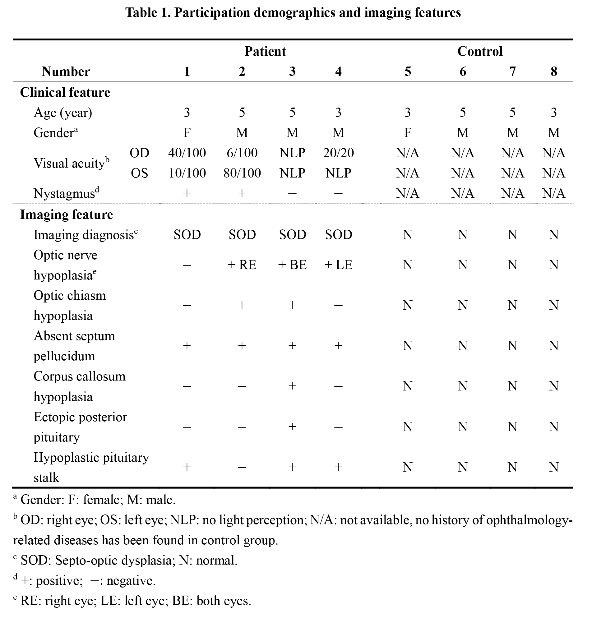
Table 1. Participation demographics and imaging features
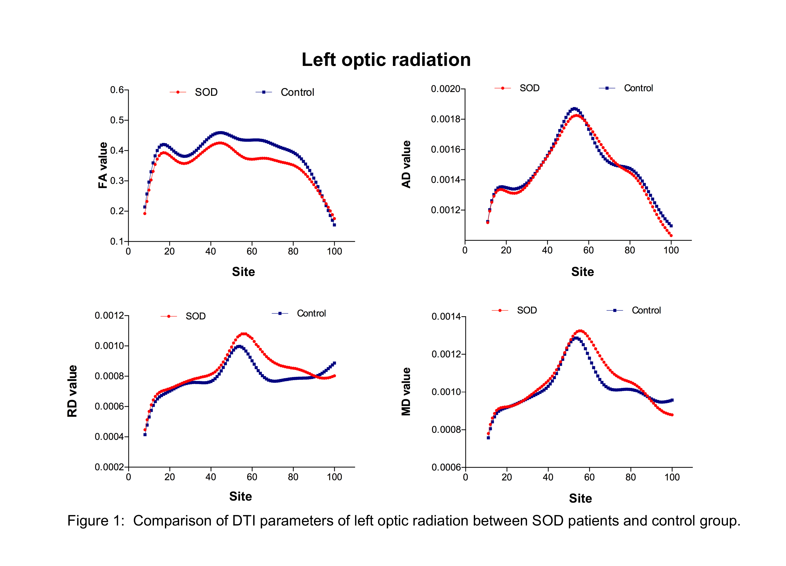
Figure 1: Comparison of DTI parameters of left optic radiation between SOD patients and control group.
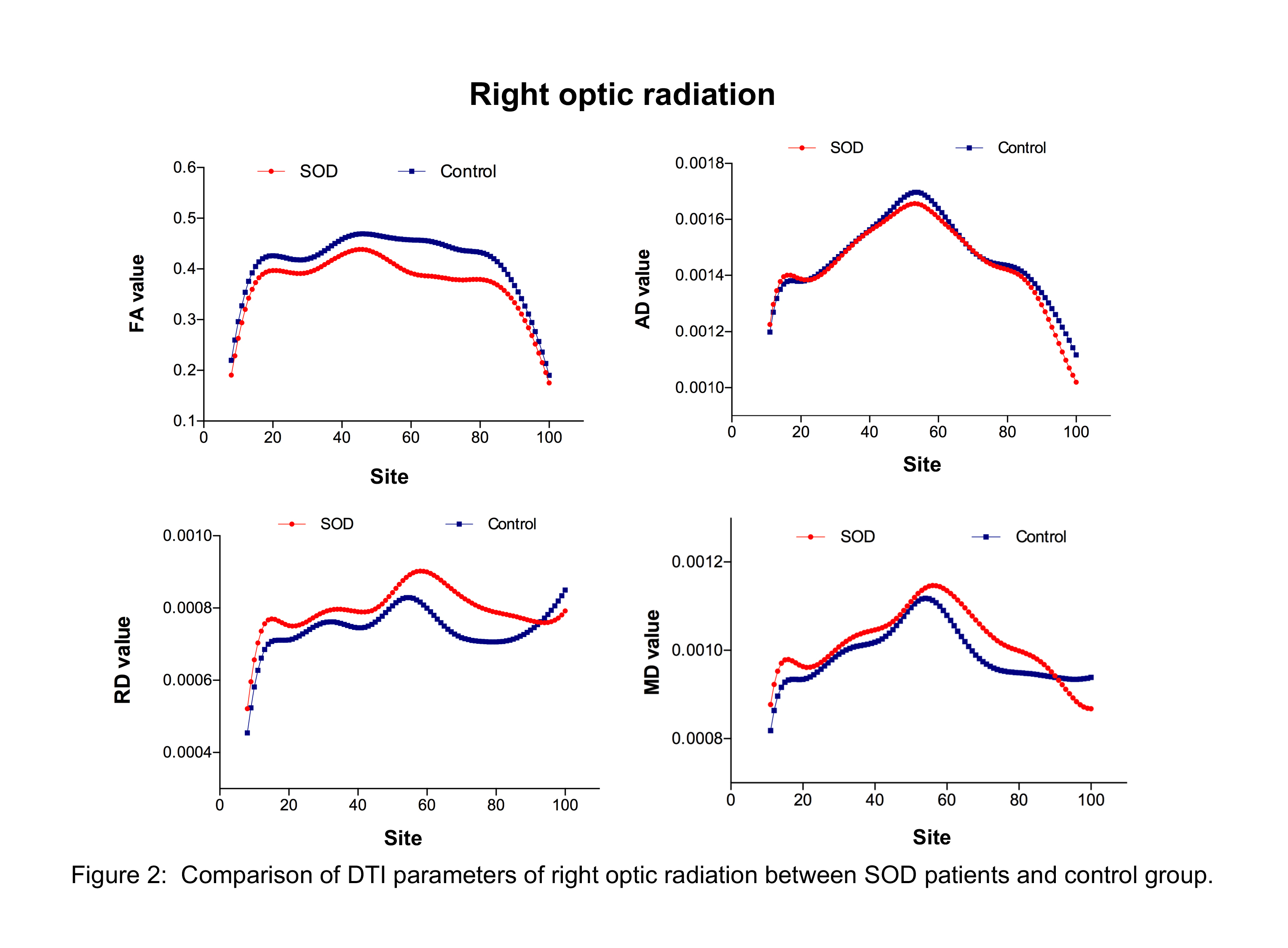
Figure 2: Comparison of DTI parameters of right optic radiation between SOD patients and control group.