4589
Investigating the brain volume and shape correlates of reading performance in children with reading difficulties1Department of Medical Imaging and Radiological Sciences, Chang Gung University, Taoyuan, Taiwan, 2Department of Special Education, National Taiwan Normal University, Taipei, Taiwan, 3Department of Psychiatry, Chang Gung Memorial Hospital, Chiayi, Taiwan, 4Medical Imaging Research Center, Radiological Research, Chang Gung University and Chang Gung Memorial Hospital, Linkou, Taoyuan, Taiwan
Synopsis
Reading disability is a special learning disorder based on the neurophysiological of the brain. Schooling children with reading disabilities (CRD) have different ways to process language from normal children (NCRD). Our study investigated the abnormality of brain volume and shape contributing to specific reading disabilities in schooling children. We found the significant difference in brain volume and shape of the parietal, temporal lobe and limbic system between CRD and NCRD. These brain areas are associated with the core phonological processing regions.
Introduction
Reading disability is a genetic disorder and the most common learning disability. Abnormalities in the cortical structure are normally observed in schooling children with reading disability (CRD) 1,2. The characteristic of CRD is slow, inaccurate reading accompanied by executive dysfunction of the brain, specifically concerning visual attention 2. Our goal was to compare the brain volumes of CRD and relatively normal children (NCRD) using voxel-based morphometry (VBM) methodology and vertex-wise shape analysis.Methods
In the study, 46 schooling children in Taipei city with the age of 10- to 11-year-old were arranged for MRI whole brain examination, including 22 children with specific reading disabilities (CRD group) and 24 sex-matched normally developing children (NCRD group). The structural T1 weighted images were obtained (TR/ TE = 2000 ms/2.98 ms, inversion time = 900 ms, FOV = 192 mm × 256 mm, spatial resolution = 1 mm × 1 mm) using a 3-Tesla MRI (MAGNETOM Prisma, Siemens, Germany) with the Magnetization Prepared Rapid Gradient Echo Imaging (MPRAGE) sequence.VBM data analysis was done using Statistical Parametric Mapping 8 (SPM8, Wellcome Department of Cognitive Neurology, London, UK) with Voxel-Based Morphometry 8 (VBM8, University of Jena, Department of Psychiatry, Jena, Germany) toolbox. Basic steps in VBM pre-processing include spatial normalization, segmentation, modulation, and smoothing. The T1 weighted images were first normalized to International Consortium for Brain Mapping (ICBM) templates, East Asian Brain and then segmented into gray matter (GM) and white matter (WM). To avoid the original difference of each brain, age, education years, and whole brain volume were used as covariates in the two-sample t-tests. A p-value of 0.01 was considered statistically significant.
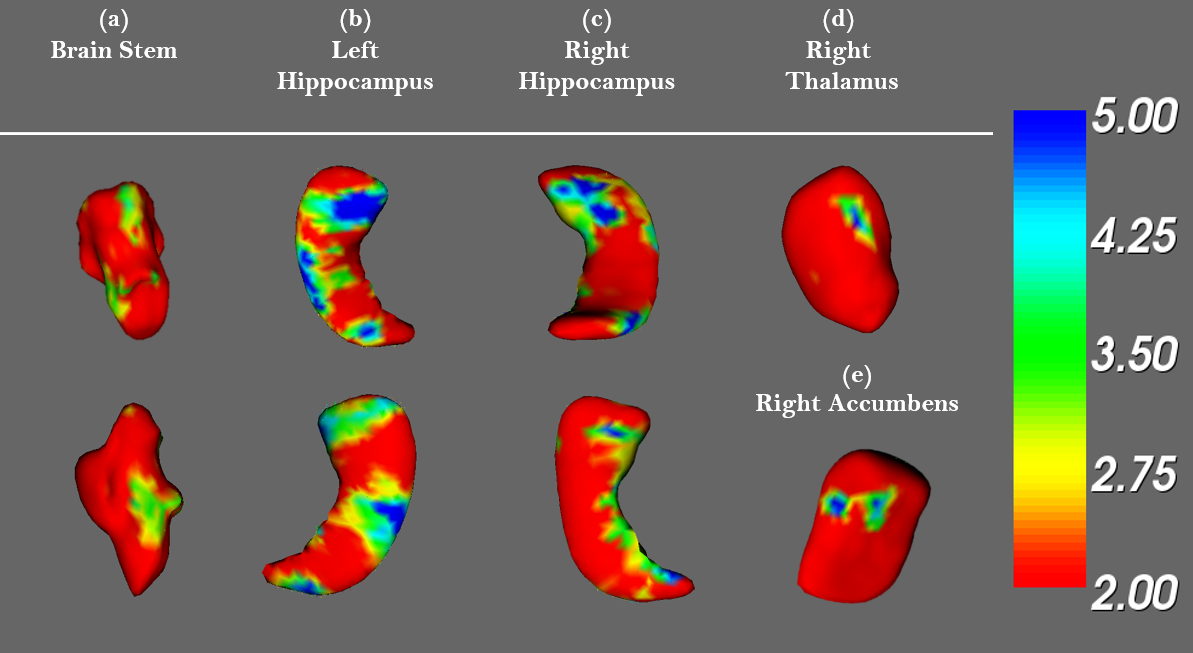
In the vertex-wise shape analysis, FMRIB Software Library (FSL, Oxford, UK) was performed to segment the brain into 15 subcortical structures, including the brain stem, bilateral hippocampus, accumbens, amygdala, caudate, pallidus, putamen, and thalamus, based on T1-weighted MRI scans using shape and appearance models. The vertex-wise analysis was then used to show the differences in the shape of subcortical structures between each group.
Results
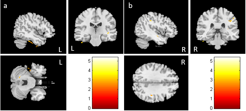
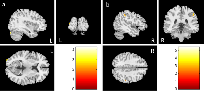
In VBM analysis, the volume of gray matter in the left fusiform gyrus and inferior parietal lobe were abnormally increased in CRD compared with NCRD (Fig. 1a & b). Moreover, the CRD group also showed a decreased volume of gray matter in the middle occipital lobe (Fig. 2a). We also observed that the volume of white matter in inferior parietal gyrus was smaller in CRD (Fig. 2b). In the shape analysis, we found the differences in the brain stem, right thalamus, right accumbens, left and right hippocampus between CRD and NCRD (Fig. 3a-e) (corrected p<0.05).Discussion
The previous study showed RD is associated with aberrant brain structure and visual function 3. Besides, we found the significant differences in brain volume of parietal, temporal lobe between CRD and NCRD. The previous study also mentioned the functional connectivity of the core phonological processing region was in the temporoparietal junction (TPJ) which was related to reading skill in an adult 4. Reduced volume of gray matter in orbitofrontal and superior temporal sulcus was relatively considered in the context of genetics studies linking to alleles that conferred risk for reading disability 5. However, their reading skills can be improved with instructional strategies 6 and music training 7.Conclusion
Firstly, RD was associated with the development of brain neurophysiological function rather than intellectual development. Secondly, CRD with abnormal brain structure demonstrated below-average reading ability and visual attention. Thirdly, our results pointed out the significant differences in brain volume and shape were consistently observed across schooling children with evidence of specific reading disability.Acknowledgements
This study was supported by the research
program MOST103-2420-H-003-008-MY3, which were sponsored by the Ministry of
Science and Technology, Taipei, Taiwan.
References
1. Handler, S. M., W. M. Fierson, O. Section on, D. Council on Children with, O. American Academy of, O. American Association for Pediatric, Strabismus and O. American Association of Certified (2011). "Learning disabilities, dyslexia, and vision." Pediatrics 127(3): e818-856.
2. Horowitz-Kraus, T. (2017). "Familial history of reading difficulty is associated with diffused bilateral brain activation during reading and greater association with visual attention abilities." Ann Dyslexia 67(3): 281-298.
3. Peterson, R. L. and B. F. Pennington (2015). "Developmental dyslexia." Annu Rev Clin Psychol 11: 283-307.
4. Achal, S., F. Hoeft and S. Bray (2016). "Individual Differences in Adult Reading Are Associated with Left Temporo-parietal to Dorsal Striatal Functional Connectivity." Cereb Cortex 26(10): 4069-4081.
5. Eckert, M. A., V. W. Berninger, K. I. Vaden, Jr., M. Gebregziabher and L. Tsu (2016). "Gray Matter Features of Reading Disability: A Combined Meta-Analytic and Direct Analysis Approach(1,2,3,4)." eNeuro 3(1).
6. Hebert, M., D. M. Kearns, J. B. Hayes, P. Bazis and S. Cooper (2018). "Why Children With Dyslexia Struggle With Writing and How to Help Them." Lang Speech Hear Serv Sch 49(4): 843-863.
7. Flaugnacco, E., L. Lopez, C. Terribili, M. Montico, S. Zoia and D. Schon (2015). "Music Training Increases Phonological Awareness and Reading Skills in Developmental Dyslexia: A Randomized Control Trial." PLoS One 10(9): e0138715.
Figures