4578
Evaluation of MRI in the Prenatal Dignosis of Cleft Lip and Palate1Shandong Unversity, Jinan, China, 2Department of Radiology, Shandong Provincial Hosptial Affiliated to Shandong University, Jinan, China, 3MR Scientific Marketing, Siemens Healthcare Ltd, Beijing, China
Synopsis
This prospective study aimed to evaluate the value of MRI in prenatal diagnosis of cleft lip and palate. Pregnant females subject with fetal facial malformations were suspected by ultrasonography (US) and then underwent 1.5 T MRI scan. The diagnosis accuracy of the prenatal US and MRI was compared with the postnatal findings. A total of 44 fetuses were followed up with lip and/or cleft palate malformation. Compared to US, MRI provides more useful information in the diagnosis of fetal cleft lip and palate,which could be used as one of the effective alternative to prenatal ultrasound diagnosis.
Introduction and Purpose
Orofacial clefts are one of the most common congenital craniofacial abnormalities 1, 2. Ultrasonography (US) is currently the first choice for antenatal diagnosis of facial malformations3. Clefts of lip and primary palate can be detected routinely, whereas clefts of the secondary palate are hard to observe, especially if they appear without any additional malformations4, 5. Besides US, magnetic resonance imaging (MRI) has been proved to be useful in prenatal examination. A common problem in most previous studies conducted to date is a selection bias that prior knowledge of US was acquired before MRI scan, thus diagnosis accuracy for fetal MRI is normally high. To avoid this bias, all MRI radiologists were blinded to the results of US in this study. Our purpose is to evaluate the accuracy of US and MRI in the diagnosis of fetal cleft lip with or without palate objectively.Methods
Patients This prospective study was approved by our institutional review board.44 Pregnant women suspected of fetal facial malformations or unable to clearly display normal maxillofacial anatomy on US from 2016 to 2019 were enrolled. The age of pregnant women was 25 ~ 35 years old, median age was 29 years old, mean age was 30 years; gestational age was 20 ~ 39 weeks, median gestational age was 28 weeks, mean gestational age 29 weeks.MRI examinations All MRI examinations were performed with a 1.5 T MAGNETOM Amira (Siemens Shenzhen Magnetic Resonance Ltd., Shenzhen, China) with 13-channel body matrix and spine array coils. All the patients were imaged in the supine position. True fast imaging with steady-state precession (True FISP) images were obtained under free-breathing condition and no maternal sedation in the axial, coronal, sagittal planes. The scan parameters were listed as follows: TR=612ms, TE= 1.93ms, FOV=380mm×380mm, 30 slices, slice thickness = 4.0 mm, 20% slice gap, acquisition matrix = 256×256, voxel size=1.3x1.3x1.3mm3, flip angle=79°, total acquisition time 19s.
Imaging analysis The MR-images were reviewed in a blinded fashion by two experienced radiologists without knowledge of indications and prior US imaging findings. If Rater 1 and Rater 2 could not reach an agreement, a third radiologist was brought into the discussion in order to reach a consensus.
Statistical analysis The prenatal US and MRI diagnoses were compared with postnatal clinical examinations after delivery or abortion. The accuracy, sensitivity and specificity of US and MRI in the diagnosis of cleft lip with or without palate were calculated based on neonatal imaging or autopsy.
Result
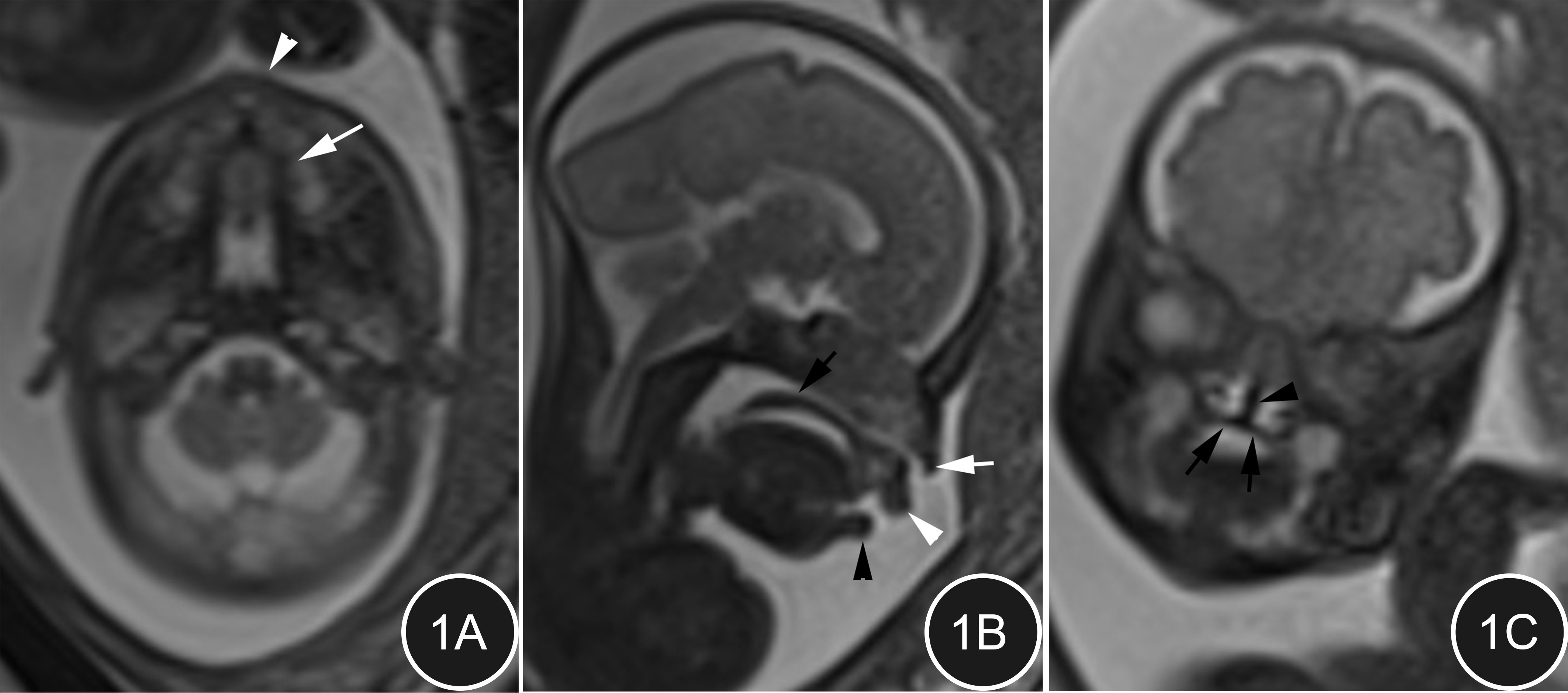
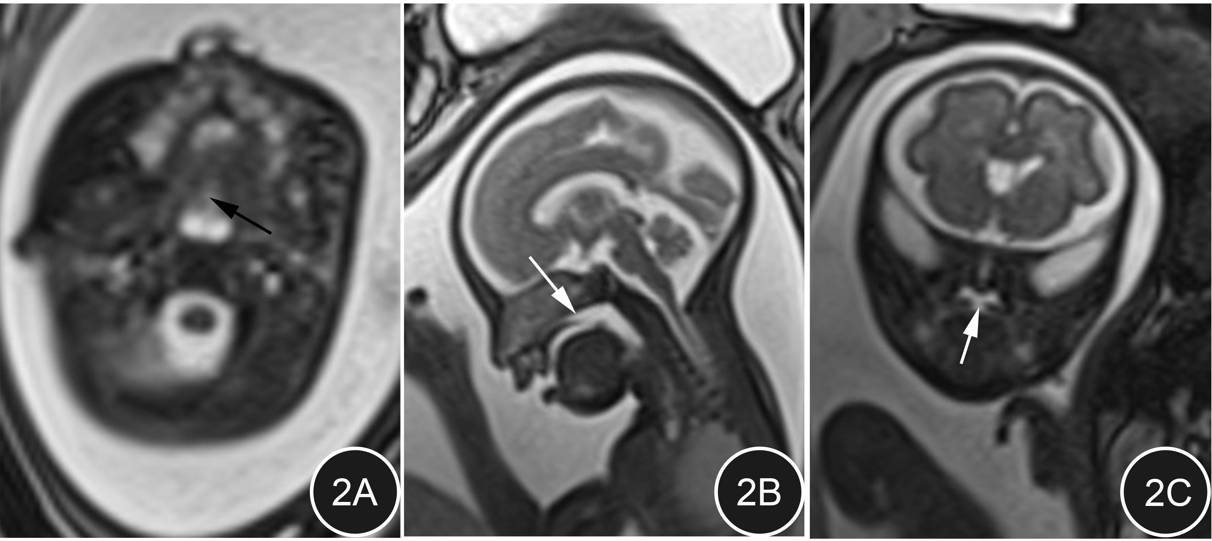
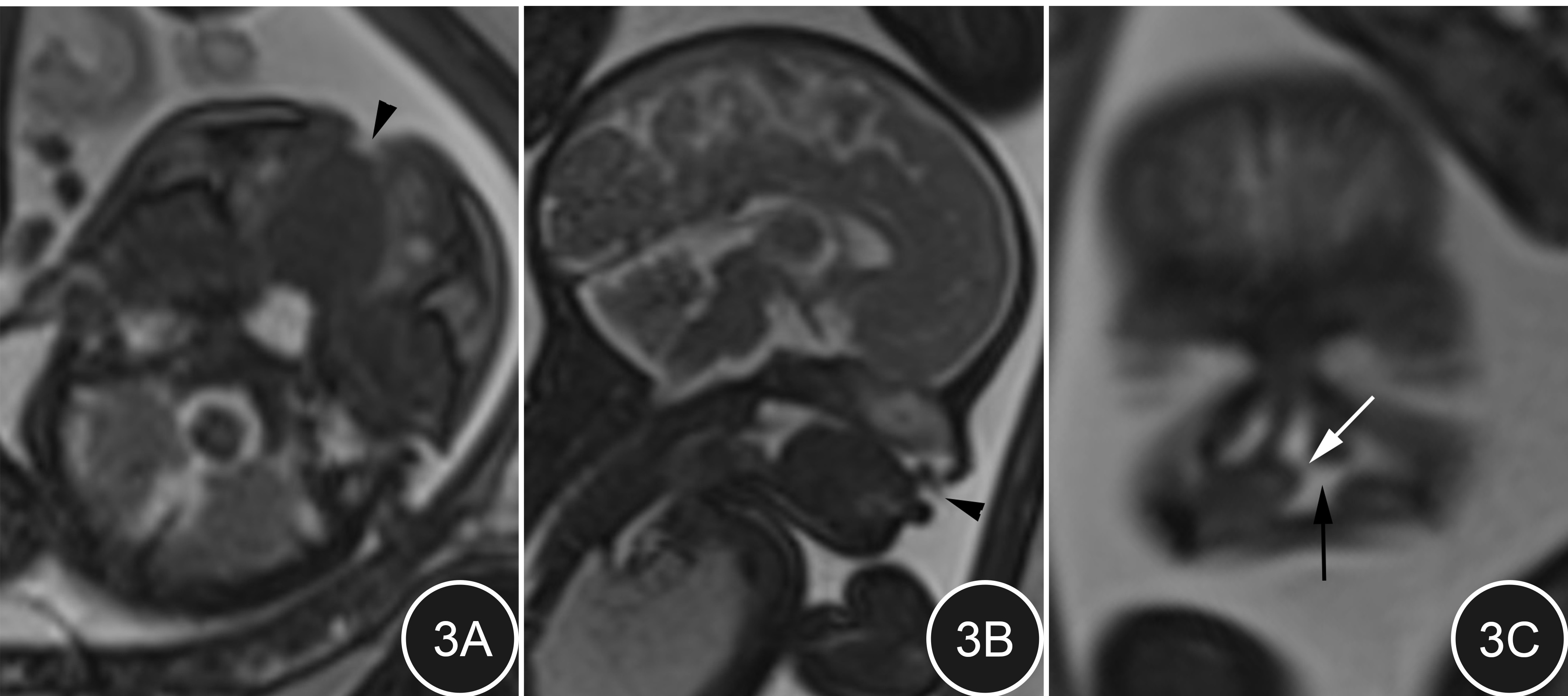
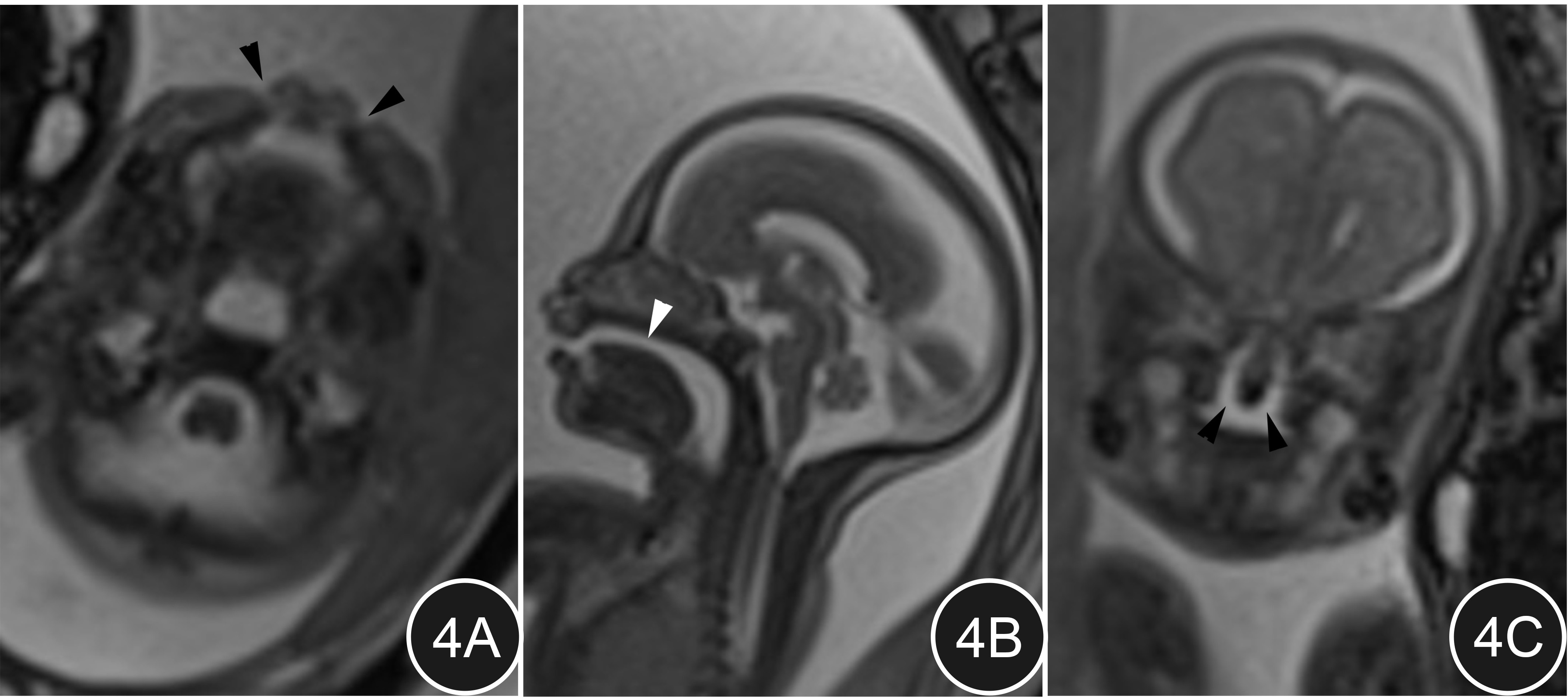
Follow-up result A total of 44 fetuses were followed up with lip and/or cleft palate malformation. The follow-up results showed unilateral isolated cleft lip in 23 cases, isolated cleft palate in 3 cases, unilateral incomplete cleft lip and cleft palate in 4 cases, unilateral complete cleft lip and cleft palate in 11 cases, and bilateral complete cleft lip accompanied with cleft palate in 3 cases.Comparison of the two modalities in displaying the extent of the cleft US clearly shows cleft lip and alveolar cleft, but it is difficult to show secondary palate cleft. MRI shows the connections between the cavities, the degree of involvement of the secondary palate and the lateral and anterior-posterior extent of the cleft better than US. MRI demonstrated the anatomy of the maxillofacial region well (figure 1A-C). It is superior to ultrasound, especially in displaying of isolated cleft palate (figure 2A-C).
Statistical result The accuracy, sensitivity and specificity in detection cleft palate was 75.0%,47.6%,100% for US and 97.7%,95.2%,100% for MRI, respectively.
Discussion and Conclusion
This is the first prospective blinded study comparing the accuracy of US and MRI in diagnosing fetal facial cleft lip and palate without ruling out multiple pregnancies and fetuses suspected associated abnormalities. Compared to US diagnosis of fetal cleft lip and palate, MRI had obvious advantages and can be used as an effective alternative to US. Especially when US had doubts about whether or not to have cleft palate, MR examination can be used as the preferred non-invasive examination method and provide more accurate information for perinatal period management and consulting.Acknowledgements
No acknowledgement found.References
1. Smarius B, Loozen C, Manten W, et al. Accurate diagnosis of prenatal cleft lip/palate by understanding the embryology[J]. World J Methodol, 2017,7(3):93-100.DOI:10.5662/wjm.v7.i3.93.
2. Clementi M, Tenconi R, Bianchi F, et al. Evaluation of prenatal diagnosis of cleft lip with or without cleft palate and cleft palate by ultrasound: experience from 20 European registries. EUROSCAN study group[J]. Prenat Diagn, 2000,20(11):870-875.
3. Loozen C S, Maarse W, Manten G T, et al. The accuracy of prenatal ultrasound in determining the type of orofacial cleft[J]. Prenat Diagn, 2015,35(7):652-655.DOI:10.1002/pd.4582.
4. Wang G, Shan R, Zhao L, et al. Fetal cleft lip with and without cleft palate: comparison between MR imaging and US for prenatal diagnosis[J]. Eur J Radiol, 2011,79(3):437-442.DOI:10.1016/j.ejrad.2010.03.026.
5. Rotten D, Levaillant J M. Two- and three-dimensional sonographic assessment of the fetal face. 2. Analysis of cleft lip, alveolus and palate[J]. Ultrasound Obstet Gynecol, 2004,24(4):402-411.DOI:10.1002/uog.1718.
Figures